3GTD image
Deposition Date 2009-03-27
Release Date 2009-07-14
Last Version Date 2023-09-06
Entry Detail
PDB ID:
3GTD
Keywords:
Title:
2.4 Angstrom crystal structure of fumarate hydratase from Rickettsia prowazekii
Biological Source:
Source Organism:
Host Organism:
Method Details:
Experimental Method:
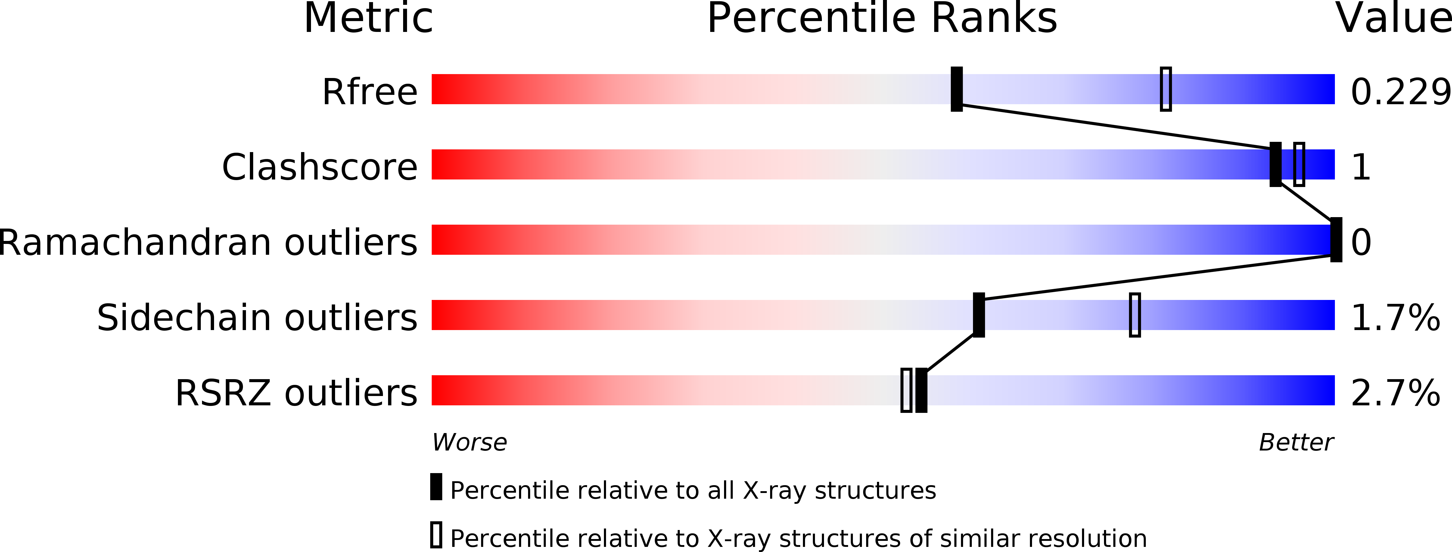
Resolution:
2.40 Å
R-Value Free:
0.22
R-Value Work:
0.19
R-Value Observed:
0.19
Space Group:
P 31 2 1
Macromolecular Entities
Polymer Type:polypeptide(L)
Molecule:Fumarate hydratase class II
Gene (Uniprot):fumC
Chain IDs:A, B
Chain Length:482
Number of Molecules:2
Biological Source:Rickettsia prowazekii
Primary Citation
Structure of fumarate hydratase from Rickettsia prowazekii, the agent of typhus and suspected relative of the mitochondria.
Acta Crystallogr.,Sect.F 67 1123 1128 (2011)
PMID: 21904061 DOI: 10.1107/S174430911102690X

Abstact

Rickettsiae are obligate intracellular parasites of eukaryotic cells that are the causative agents responsible for spotted fever and typhus. Their small genome (about 800 protein-coding genes) is highly conserved across species and has been postulated as the ancestor of the mitochondria. No genes that are required for glycolysis are found in the Rickettsia prowazekii or mitochondrial genomes, but a complete set of genes encoding components of the tricarboxylic acid cycle and the respiratory-chain complex is found in both. A 2.4 Å resolution crystal structure of R. prowazekii fumarate hydratase, an enzyme catalyzing the third step of the tricarboxylic acid cycle pathway that ultimately converts phosphoenolpyruvate into succinyl-CoA, has been solved. A structure alignment with human mitochondrial fumarate hydratase highlights the close similarity between R. prowazekii and mitochondrial enzymes.

Legend

Protein

Chemical

Disease

Primary Citation of related structures