3GR1 image
Deposition Date 2009-03-24
Release Date 2009-05-19
Last Version Date 2023-09-06
Entry Detail
PDB ID:
3GR1
Title:
Periplasmic domain of the T3SS inner membrane protein PrgH from S.typhimurium (fragment 170-392)
Biological Source:
Source Organism:
Host Organism:
Method Details:
Experimental Method:
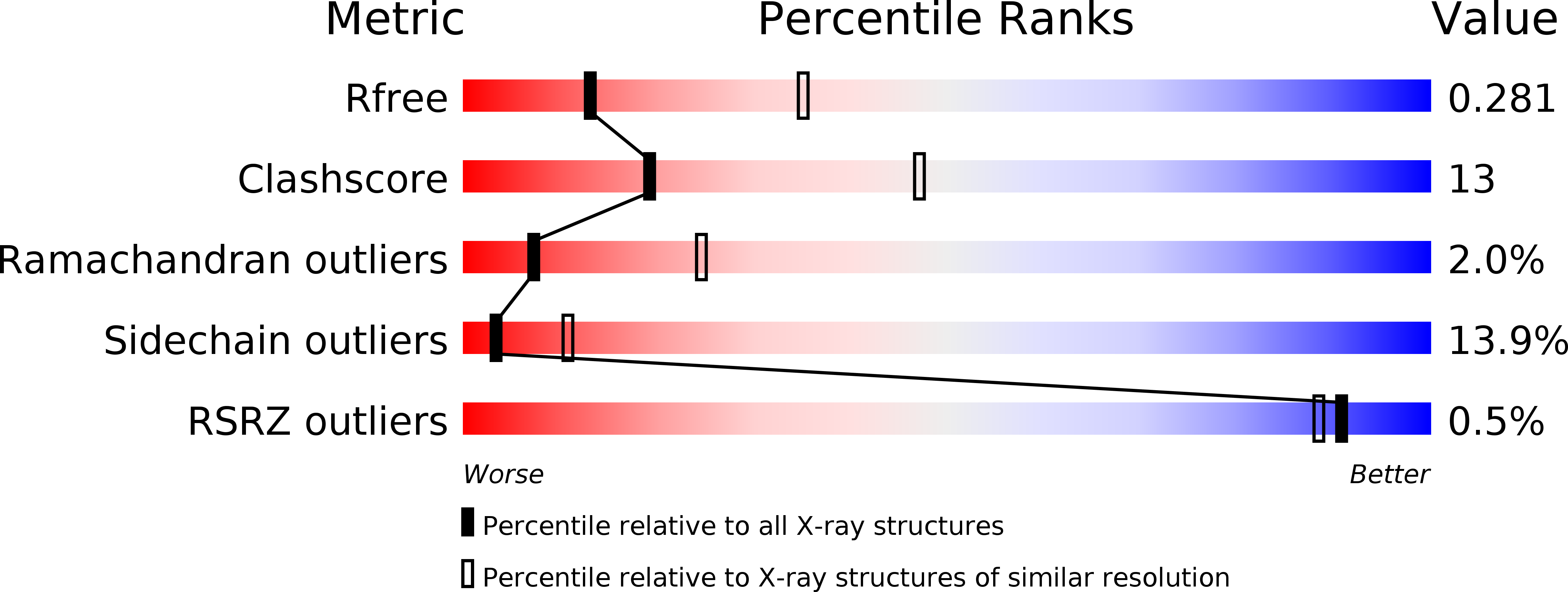
Resolution:
2.80 Å
R-Value Free:
0.29
R-Value Work:
0.24
Space Group:
C 2 2 21
Macromolecular Entities
Polymer Type:polypeptide(L)
Molecule:Protein prgH
Gene (Uniprot):prgH
Chain IDs:A, B, C, D, E, F, G, H
Chain Length:227
Number of Molecules:8
Biological Source:Salmonella typhimurium
Ligand Molecules
Primary Citation
A conserved structural motif mediates formation of the periplasmic rings in the type III secretion system.
Nat.Struct.Mol.Biol. 16 468 476 (2009)
PMID: 19396170 DOI: 10.1038/nsmb.1603

Abstact

The type III secretion system (T3SS) is a macromolecular 'injectisome' that allows bacterial pathogens to transport virulence proteins into the eukaryotic host cell. This macromolecular complex is composed of connected ring-like structures that span both bacterial membranes. The crystal structures of the periplasmic domain of the outer membrane secretin EscC and the inner membrane protein PrgH reveal the conservation of a modular fold among the three proteins that form the outer membrane and inner membrane rings of the T3SS. This leads to the hypothesis that this conserved fold provides a common ring-building motif that allows for the assembly of the variably sized outer membrane and inner membrane rings characteristic of the T3SS. Using an integrated structural and experimental approach, we generated ring models for the periplasmic domain of EscC and placed them in the context of the assembled T3SS, providing evidence for direct interaction between the outer membrane and inner membrane ring components and an unprecedented span of the outer membrane secretin.

Legend

Protein

Chemical

Disease

Primary Citation of related structures