Deposition Date
2009-03-24
Release Date
2009-06-02
Last Version Date
2024-02-21
Entry Detail
PDB ID:
3GQJ
Keywords:
Title:
Crystal structure of Cell Inhibiting Factor (Cif) from Photorhabdus luminescens
Biological Source:
Source Organism(s):
Photorhabdus luminescens subsp. laumondii (Taxon ID: 141679)
Expression System(s):
Method Details:
Experimental Method:
Resolution:
1.85 Å
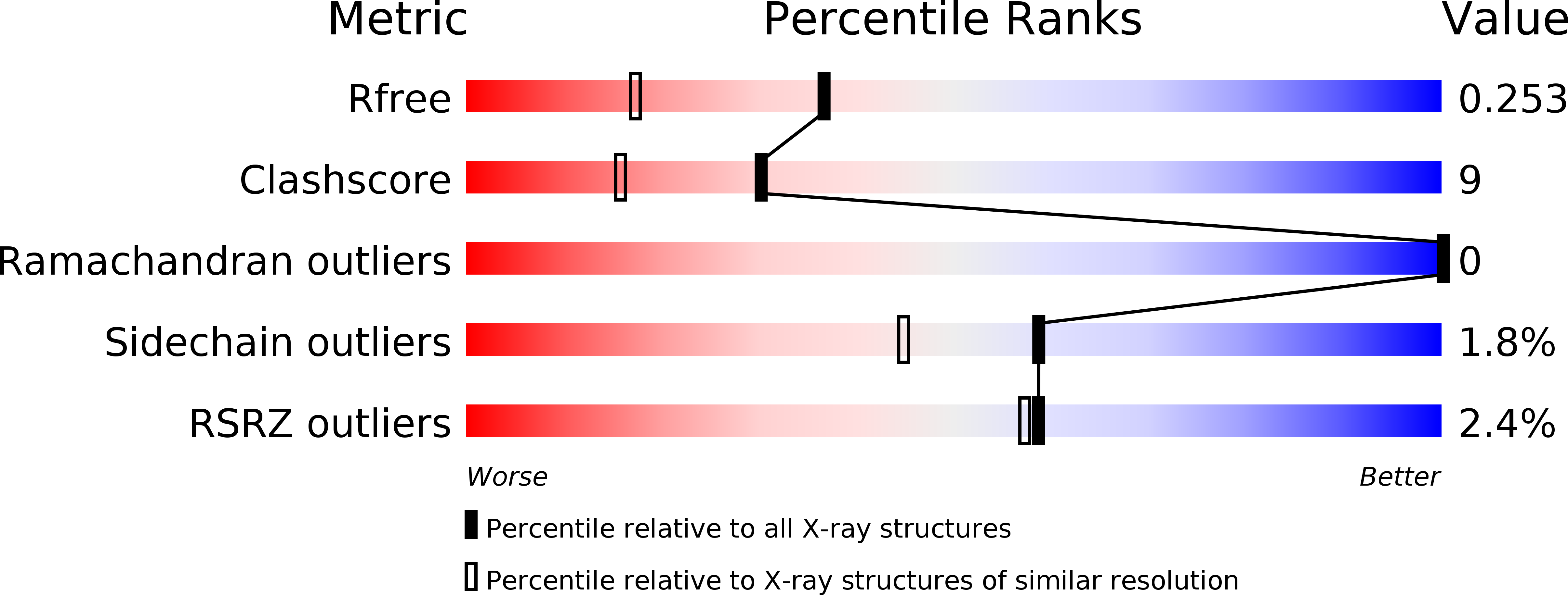
R-Value Free:
0.27
R-Value Work:
0.22
R-Value Observed:
0.22
Space Group:
P 61 2 2