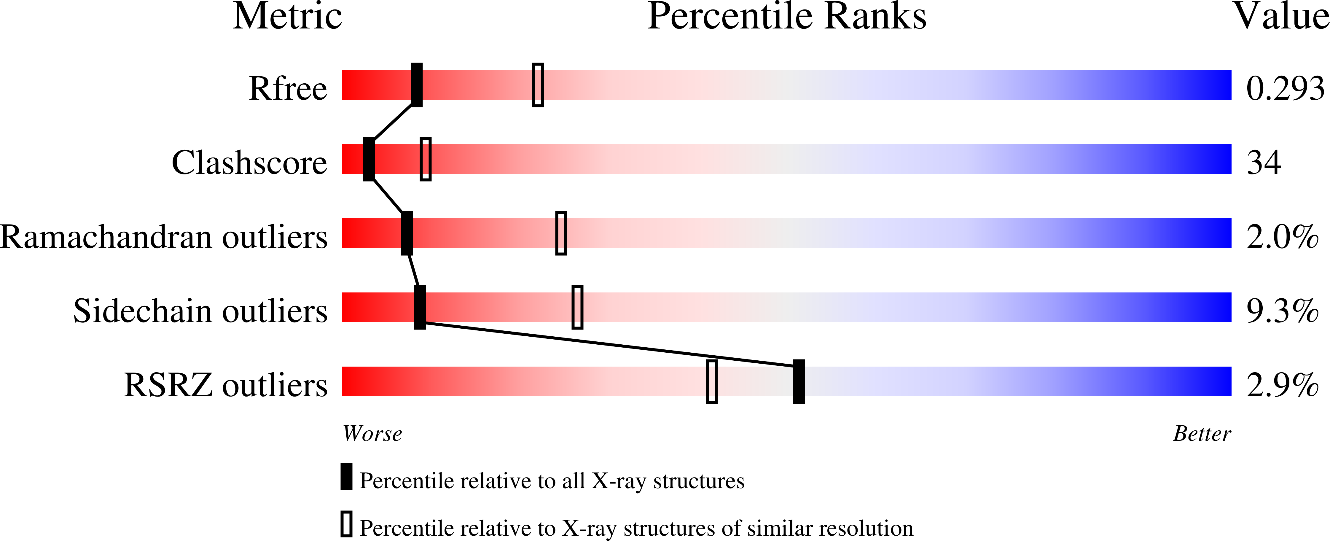
3GQB image
Deposition Date 2009-03-24
Release Date 2009-11-24
Last Version Date 2024-02-21
Entry Detail
PDB ID:
3GQB
Keywords:
Title:
Crystal Structure of the A3B3 complex from V-ATPase
Biological Source:
Source Organism:
Host Organism:
Method Details:
Experimental Method:
Resolution:
2.80 Å
R-Value Free:
0.28
R-Value Work:
0.24
R-Value Observed:
0.25
Space Group:
P 3 2 1
Macromolecular Entities
Structures with similar UniProt ID
Protein Blast
Polymer Type:polypeptide(L)
Molecule:V-type ATP synthase alpha chain
Gene (Uniprot):atpA
Mutagens:C28S, T235S, C255S, C507S
Chain IDs:A, C
Chain Length:578
Number of Molecules:2
Biological Source:Thermus thermophilus HB8
Structures with similar UniProt ID
Protein Blast
Polymer Type:polypeptide(L)
Molecule:V-type ATP synthase beta chain
Gene (Uniprot):atpB
Mutagens:C264S
Chain IDs:B, D
Chain Length:464
Number of Molecules:2
Biological Source:Thermus thermophilus HB8
Primary Citation
Crystal structure of A(3)B(3) complex of V-ATPase from Thermus thermophilus.
Embo J. 28 3771 3779 (2009)
PMID: 19893485 DOI: 10.1038/emboj.2009.310

Abstact

Vacuolar-type ATPases (V-ATPases) exist in various cellular membranes of many organisms to regulate physiological processes by controlling the acidic environment. Here, we have determined the crystal structure of the A(3)B(3) subcomplex of V-ATPase at 2.8 A resolution. The overall construction of the A(3)B(3) subcomplex is significantly different from that of the alpha(3)beta(3) sub-domain in F(o)F(1)-ATP synthase, because of the presence of a protruding 'bulge' domain feature in the catalytic A subunits. The A(3)B(3) subcomplex structure provides the first molecular insight at the catalytic and non-catalytic interfaces, which was not possible in the structures of the separate subunits alone. Specifically, in the non-catalytic interface, the B subunit seems to be incapable of binding ATP, which is a marked difference from the situation indicated by the structure of the F(o)F(1)-ATP synthase. In the catalytic interface, our mutational analysis, on the basis of the A(3)B(3) structure, has highlighted the presence of a cluster composed of key hydrophobic residues, which are essential for ATP hydrolysis by V-ATPases.

Legend

Protein

Chemical

Disease

Primary Citation of related structures