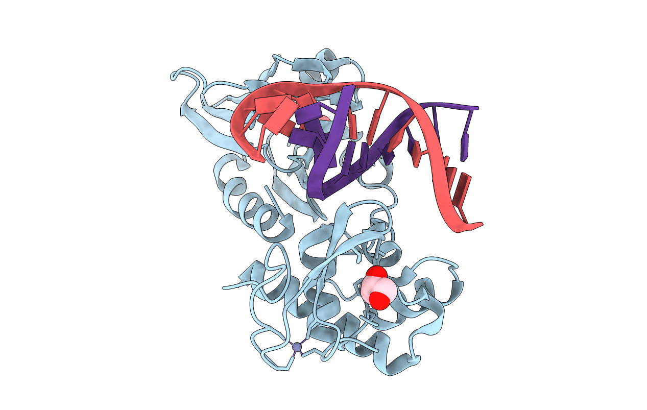
3GQ5 image
Deposition Date 2009-03-23
Release Date 2009-11-10
Last Version Date 2024-02-21
Entry Detail
PDB ID:
3GQ5
Keywords:
Title:
Sequence-matched MutM Interrogation Complex 5 (IC5)
Biological Source:
Source Organism:
Host Organism:
Method Details:
Experimental Method:
Resolution:
1.90 Å
R-Value Free:
0.22
R-Value Work:
0.20
R-Value Observed:
0.20
Space Group:
P 21 21 21
Macromolecular Entities
Polymer Type:polypeptide(L)
Molecule:DNA glycosylase
Gene (Uniprot):mutM
Mutations:Q166C, T224P
Chain IDs:A
Chain Length:273
Number of Molecules:1
Biological Source:Geobacillus stearothermophilus
Primary Citation
Encounter and extrusion of an intrahelical lesion by a DNA repair enzyme.
Nature 462 762 766 (2009)
PMID: 20010681 DOI: 10.1038/nature08561

Abstact

How living systems detect the presence of genotoxic damage embedded in a million-fold excess of undamaged DNA is an unresolved question in biology. Here we have captured and structurally elucidated a base-excision DNA repair enzyme, MutM, at the stage of initial encounter with a damaged nucleobase, 8-oxoguanine (oxoG), nested within a DNA duplex. Three structures of intrahelical oxoG-encounter complexes are compared with sequence-matched structures containing a normal G base in place of an oxoG lesion. Although the protein-DNA interfaces in the matched complexes differ by only two atoms-those that distinguish oxoG from G-their pronounced structural differences indicate that MutM can detect a lesion in DNA even at the earliest stages of encounter. All-atom computer simulations show the pathway by which encounter of the enzyme with the lesion causes extrusion from the DNA duplex, and they elucidate the critical free energy difference between oxoG and G along the extrusion pathway.

Legend

Protein

Chemical

Disease

Primary Citation of related structures