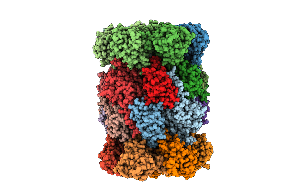
3GPW image
Entry Detail
PDB ID:
3GPW
Keywords:
Title:
Crystal structure of the yeast 20S proteasome in complex with Salinosporamide derivatives: irreversible inhibitor ligand
Biological Source:
Source Organism:
PDB Version:
Deposition Date:
2009-03-23
Release Date:
2009-09-15
Method Details:
Experimental Method:
Resolution:
2.50 Å
R-Value Free:
0.25
R-Value Work:
0.23
R-Value Observed:
0.23
Space Group:
P 1 21 1
Macromolecular Entities
Polymer Type:polypeptide(L)
Description:Proteasome component Y7
Chain IDs:A, O
Chain Length:250
Number of Molecules:2
Biological Source:Saccharomyces cerevisiae
Polymer Type:polypeptide(L)
Description:Proteasome component Y13
Chain IDs:B, P
Chain Length:244
Number of Molecules:2
Biological Source:Saccharomyces cerevisiae
Polymer Type:polypeptide(L)
Description:Proteasome component PRE6
Chain IDs:C, Q
Chain Length:241
Number of Molecules:2
Biological Source:Saccharomyces cerevisiae
Polymer Type:polypeptide(L)
Description:Proteasome component PUP2
Chain IDs:D, R
Chain Length:242
Number of Molecules:2
Biological Source:Saccharomyces cerevisiae
Polymer Type:polypeptide(L)
Description:Proteasome component PRE5
Chain IDs:E, S
Chain Length:233
Number of Molecules:2
Biological Source:Saccharomyces cerevisiae
Polymer Type:polypeptide(L)
Description:Proteasome component C1
Chain IDs:F, T
Chain Length:244
Number of Molecules:2
Biological Source:Saccharomyces cerevisiae
Polymer Type:polypeptide(L)
Description:Proteasome component C7-alpha
Chain IDs:G, U
Chain Length:243
Number of Molecules:2
Biological Source:Saccharomyces cerevisiae
Polymer Type:polypeptide(L)
Description:Proteasome component PUP1
Chain IDs:H, V
Chain Length:222
Number of Molecules:2
Biological Source:Saccharomyces cerevisiae
Polymer Type:polypeptide(L)
Description:Proteasome component PUP3
Chain IDs:I, W
Chain Length:204
Number of Molecules:2
Biological Source:Saccharomyces cerevisiae
Polymer Type:polypeptide(L)
Description:Proteasome component C11
Chain IDs:J, X
Chain Length:198
Number of Molecules:2
Biological Source:Saccharomyces cerevisiae
Polymer Type:polypeptide(L)
Description:Proteasome component PRE2
Chain IDs:K, Y
Chain Length:212
Number of Molecules:2
Biological Source:Saccharomyces cerevisiae
Polymer Type:polypeptide(L)
Description:Proteasome component C5
Chain IDs:L, Z
Chain Length:222
Number of Molecules:2
Biological Source:Saccharomyces cerevisiae
Polymer Type:polypeptide(L)
Description:Proteasome component PRE4
Chain IDs:M, AA (auth: 1)
Chain Length:233
Number of Molecules:2
Biological Source:Saccharomyces cerevisiae
Polymer Type:polypeptide(L)
Description:Proteasome component PRE3
Chain IDs:N, BA (auth: 2)
Chain Length:196
Number of Molecules:2
Biological Source:Saccharomyces cerevisiae
Ligand Molecules
Primary Citation
Snapshots of the fluorosalinosporamide/20S complex offer mechanistic insights for fine tuning proteasome inhibition
J.Med.Chem. 52 5420 5428 (2009)
PMID: 19678642 DOI: 10.1021/jm900559x

Abstact

Many marketed drugs contain fluorine, reflecting its ability to modulate a variety of biological responses. The unique 20S proteasome inhibition profile of fluorosalinosporamide compared to chlorinated anticancer agent salinosporamide A (NPI-0052) is exemplary and relates to each halogen's leaving group potential. Crystal structures of fluoro-, hydroxy-, and bromosalinosporamide in complex with the yeast 20S proteasome core particle (CP) provide mechanistic insights into ligand binding and leaving group elimination and the ability to fine-tune the duration of proteasome inhibition. Fluorosalinosporamide/CP crystal structures determined over time offer striking snapshots of the ligand trapped with an intact fluoroethyl group in anticipation of fluoride elimination, followed by complete nucleophilic displacement of fluoride to give the highly stabilized cyclic ether found for salinosporamide A and bromosalinosporamide. This two-step reaction pathway is consistent with a mechanism for partially reversible proteasome inhibition by fluorosalinosporamide. Proteasome catalyzed fluoride displacement provides preliminary insights into the active site Thr1N pK(a).

Legend

Protein

Chemical

Disease

Primary Citation of related structures