Deposition Date
2009-03-23
Release Date
2009-05-05
Last Version Date
2024-04-03
Entry Detail
PDB ID:
3GPE
Keywords:
Title:
Crystal Structure Analysis of PKC (alpha)-C2 domain complexed with Ca2+ and PtdIns(4,5)P2
Biological Source:
Source Organism(s):
Rattus norvegicus (Taxon ID: 10116)
Expression System(s):
Method Details:
Experimental Method:
Resolution:
2.00 Å
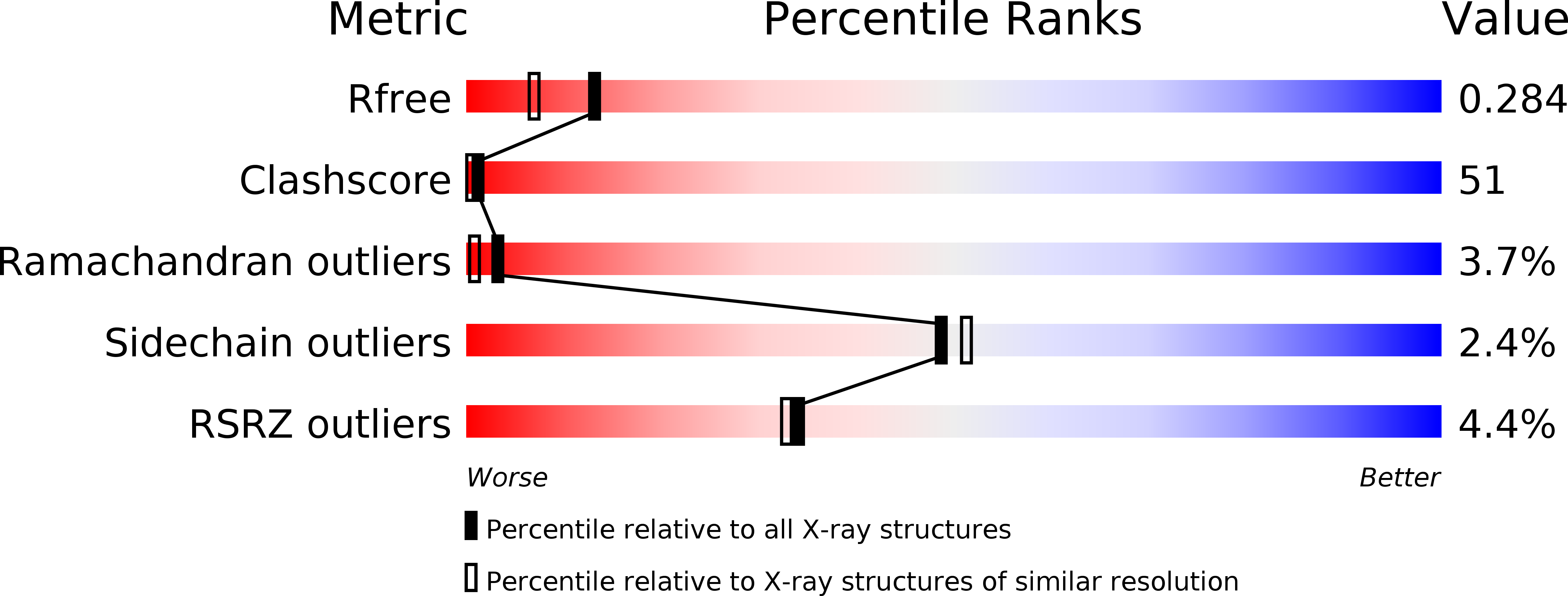
R-Value Free:
0.27
R-Value Work:
0.24
R-Value Observed:
0.24
Space Group:
P 32 2 1