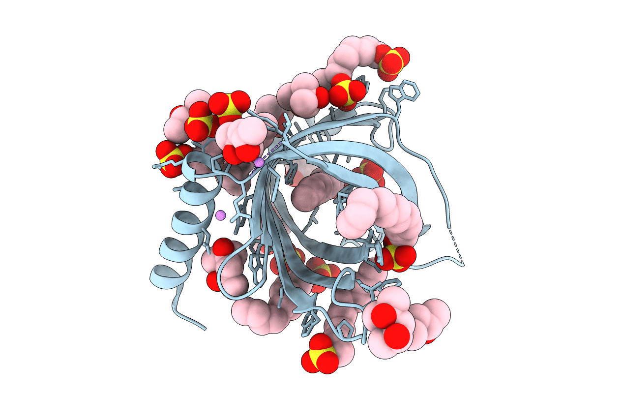
3GP6 image
Deposition Date 2009-03-20
Release Date 2010-06-23
Last Version Date 2023-09-06
Entry Detail
PDB ID:
3GP6
Keywords:
Title:
Crystal structure of PagP in SDS/MPD
Biological Source:
Source Organism:
Host Organism:
Method Details:
Experimental Method:
Resolution:
1.40 Å
R-Value Free:
0.20
R-Value Work:
0.17
R-Value Observed:
0.17
Space Group:
P 62 2 2
Macromolecular Entities
Polymer Type:polypeptide(L)
Molecule:Protein pagP
Gene (Uniprot):pagP
Chain IDs:A
Chain Length:163
Number of Molecules:1
Biological Source:Escherichia coli
Primary Citation
PagP crystallized from SDS/cosolvent reveals the route for phospholipid access to the hydrocarbon ruler.
Structure 18 1210 1219 (2010)
PMID: 20826347 DOI: 10.1016/j.str.2010.06.014

Abstact

Enzymatic reactions involving bilayer lipids occur in an environment with strict physical and topological constraints. The integral membrane enzyme PagP transfers a palmitoyl group from a phospholipid to lipid A in order to assist Escherichia coli in evading host immune defenses during infection. PagP measures the palmitoyl group with an internal hydrocarbon ruler that is formed in the interior of the eight-stranded antiparallel β barrel. The access and egress of the palmitoyl group is thought to take a lateral route from the bilayer phase to the barrel interior. Molecular dynamics, mutagenesis, and a 1.4 A crystal structure of PagP in an SDS / 2-methyl-2,4-pentanediol (MPD) cosolvent system reveal that phospholipid access occurs at the crenel present between strands F and G of PagP. In this way, the phospholipid head group can remain exposed to the cell exterior while the lipid acyl chain remains in a predominantly hydrophobic environment as it translocates to the protein interior.

Legend

Protein

Chemical

Disease

Primary Citation of related structures