3GOR image
Deposition Date 2009-03-19
Release Date 2009-05-19
Last Version Date 2024-10-30
Entry Detail
PDB ID:
3GOR
Title:
Crystal structure of putative metal-dependent hydrolase APC36150
Biological Source:
Source Organism:
Host Organism:
Method Details:
Experimental Method:
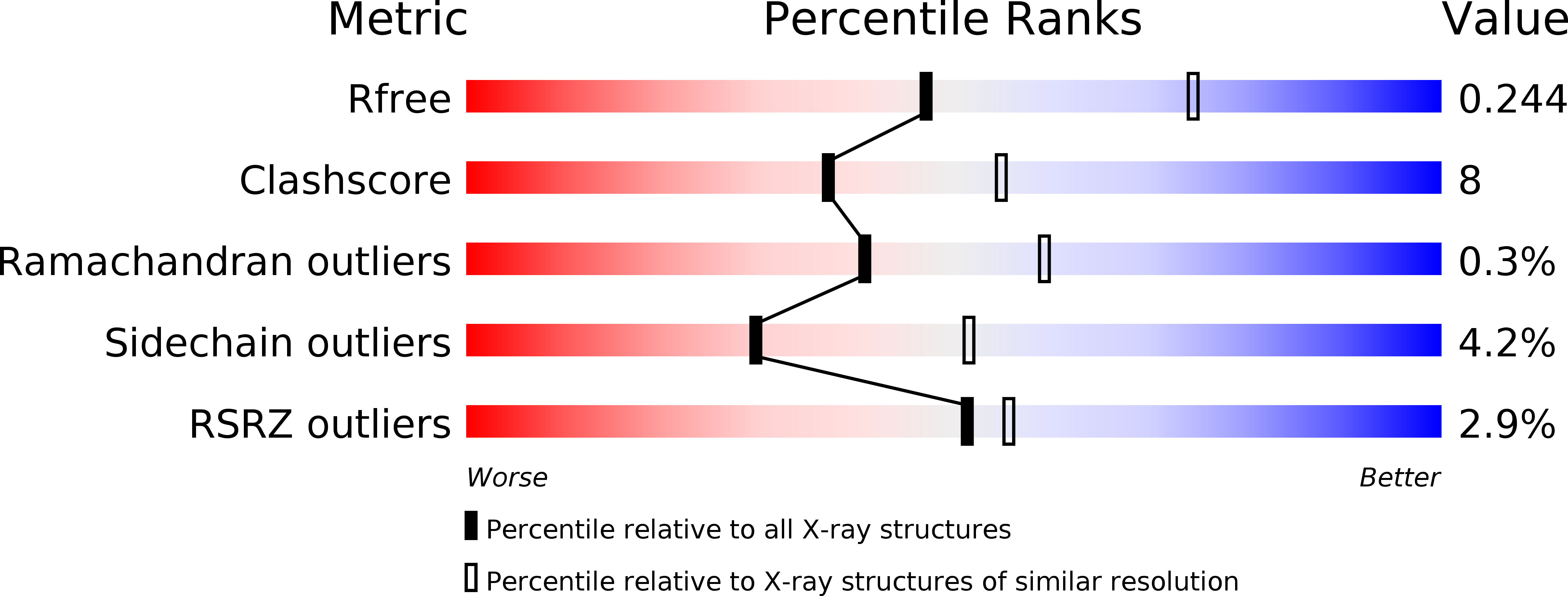
Resolution:
2.51 Å
R-Value Free:
0.24
R-Value Work:
0.18
R-Value Observed:
0.18
Space Group:
P 21 21 21
Macromolecular Entities
Polymer Type:polypeptide(L)
Molecule:Putative metal-dependent hydrolase
Chain IDs:A, B, C, D
Chain Length:157
Number of Molecules:4
Biological Source:Geobacillus stearothermophilus
Modified Residue
Compound ID Chain ID Parent Comp ID Details 2D Image
MSE A MET SELENOMETHIONINE
Ligand Molecules
Primary Citation
The structure of DinB from Geobacillus stearothermophilus: a representative of a unique four-helix-bundle superfamily.
Acta Crystallogr.,Sect.F 66 219 224 (2010)
PMID: 20208147 DOI: 10.1107/S1744309109053913

Abstact

The crystal structure of the dinB gene product from Geobacillus stearothermophilus (GsDinB) is reported at 2.5 A resolution. The dinB gene is one of the DNA-damage-induced genes and the corresponding protein, DinB, is the founding member of a Pfam family with no known function. The protein contains a four-helix up-down-down-up bundle that has previously been described in the literature in three disparate proteins: the enzyme MDMPI (mycothiol-dependent maleylpyruvate isomerase), YfiT and TTHA0303, a member of a small DUF (domain of unknown function). However, a search of the DALI structural database revealed similarities to a further 11 new unpublished structures contributed by structural genomics centers. The sequences of these proteins are quite divergent and represent several Pfam families, yet their structures are quite similar and most (but not all) seem to have the ability to coordinate a metal ion using a conserved histidine-triad motif. The structural similarities of these diverse proteins suggest that a new Pfam clan encompassing the families that share this fold should be created. The proteins that share this fold exhibit four different quaternary structures: monomeric and three different dimeric forms.

Legend

Protein

Chemical

Disease

Primary Citation of related structures