Deposition Date
2009-03-19
Release Date
2009-07-07
Last Version Date
2023-09-06
Entry Detail
PDB ID:
3GOP
Keywords:
Title:
Crystal structure of the EGF receptor juxtamembrane and kinase domains
Biological Source:
Source Organism(s):
Homo sapiens (Taxon ID: 9606)
Expression System(s):
Method Details:
Experimental Method:
Resolution:
2.80 Å
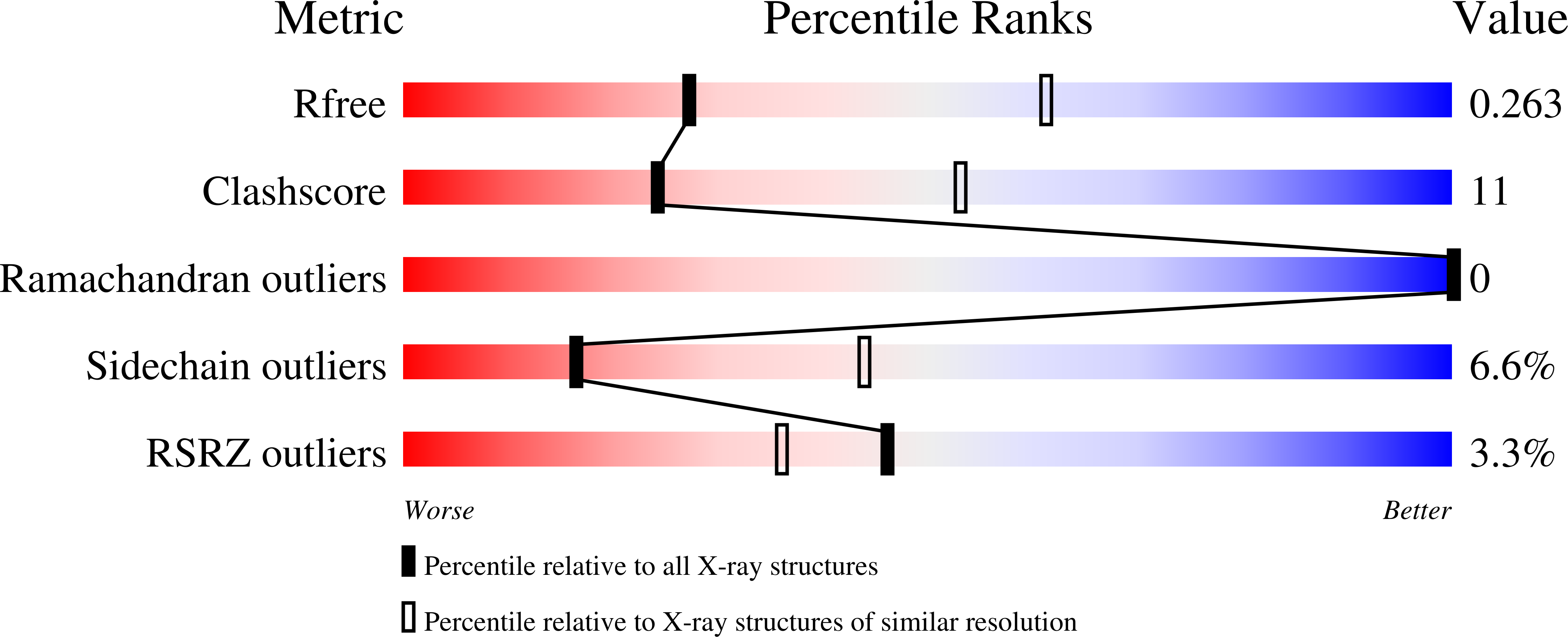
R-Value Free:
0.25
R-Value Work:
0.20
R-Value Observed:
0.21
Space Group:
P 43 21 2