Deposition Date
2009-03-15
Release Date
2009-06-02
Last Version Date
2024-11-06
Entry Detail
PDB ID:
3GMT
Keywords:
Title:
Crystal structure of adenylate kinase from burkholderia pseudomallei
Biological Source:
Source Organism(s):
Burkholderia pseudomallei 1710b (Taxon ID: 320372)
Expression System(s):
Method Details:
Experimental Method:
Resolution:
2.10 Å
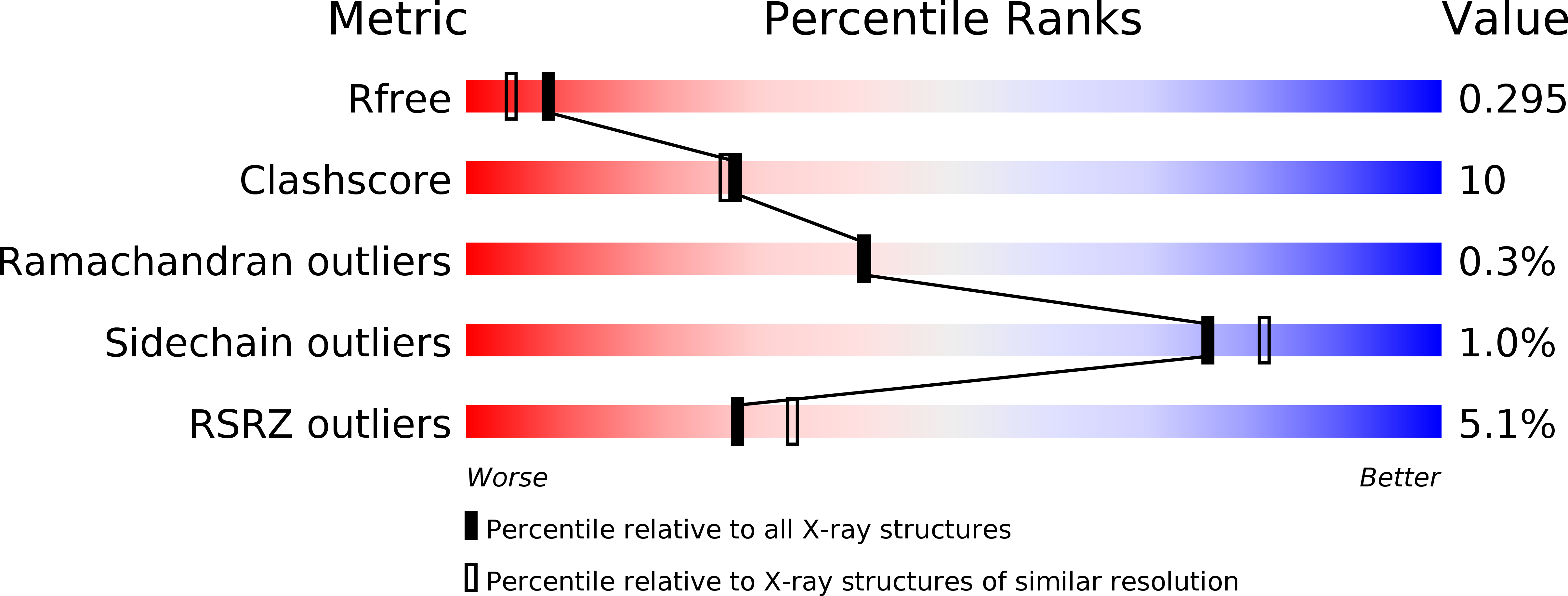
R-Value Free:
0.29
R-Value Work:
0.23
R-Value Observed:
0.23
Space Group:
P 1 21 1