3GMA image
Entry Detail
PDB ID:
3GMA
Keywords:
Title:
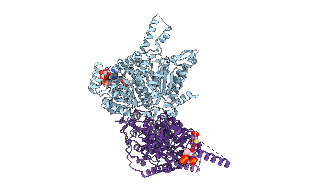
Glutaconyl-coA decarboxylase A subunit from Clostridium symbiosum co-crystallized with glutaryl-CoA
Biological Source:
Source Organism:
Host Organism:
PDB Version:
Deposition Date:
2009-03-13
Release Date:
2009-07-28
Method Details:
Experimental Method:
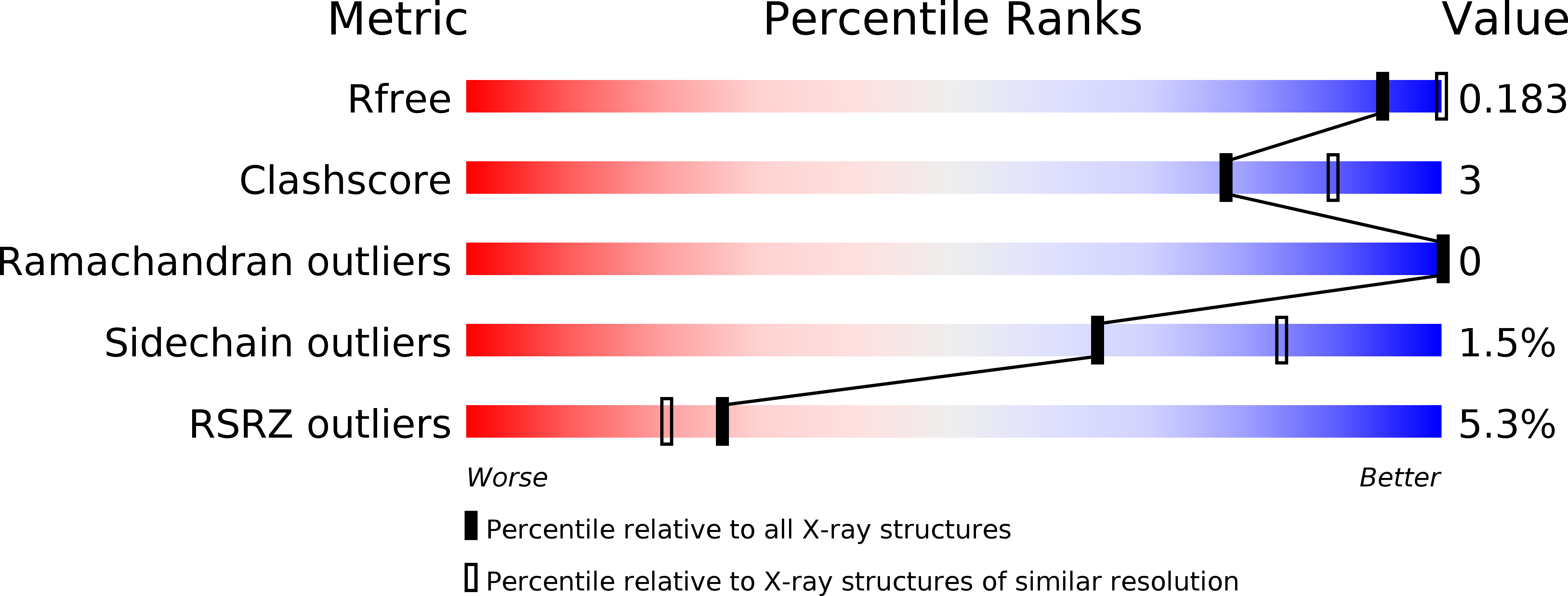
Resolution:
2.60 Å
R-Value Free:
0.20
R-Value Work:
0.18
R-Value Observed:
0.18
Space Group:
P 31 2 1
Macromolecular Entities
Polymer Type:polypeptide(L)
Description:Glutaconyl-CoA decarboxylase subunit A
Chain IDs:A, B
Chain Length:588
Number of Molecules:2
Biological Source:Clostridium symbiosum
Ligand Molecules
Primary Citation
An asymmetric model for Na+-translocating glutaconyl-CoA decarboxylases
J.Biol.Chem. 284 28401 28409 (2009)
PMID: 19654317 DOI: 10.1074/jbc.M109.037762

Abstact

Glutaconyl-CoA decarboxylase (Gcd) couples the biotin-dependent decarboxylation of glutaconyl-CoA with the generation of an electrochemical Na(+) gradient. Sequencing of the genes encoding all subunits of the Clostridium symbiosum decarboxylase membrane complex revealed that it comprises two distinct biotin carrier subunits, GcdC(1) and GcdC(2), which differ in the length of a central alanine- and proline-rich linker domain. Co-crystallization of the decarboxylase subunit GcdA with the substrate glutaconyl-CoA, the product crotonyl-CoA, and the substrate analogue glutaryl-CoA, respectively, resulted in a high resolution model for substrate binding and catalysis revealing remarkable structural changes upon substrate binding. Unlike the GcdA structure from Acidaminococcus fermentans, these data suggest that in intact Gcd complexes, GcdA is associated as a tetramer crisscrossed by a network of solvent-filled tunnels.

Legend

Protein

Chemical

Disease

Primary Citation of related structures