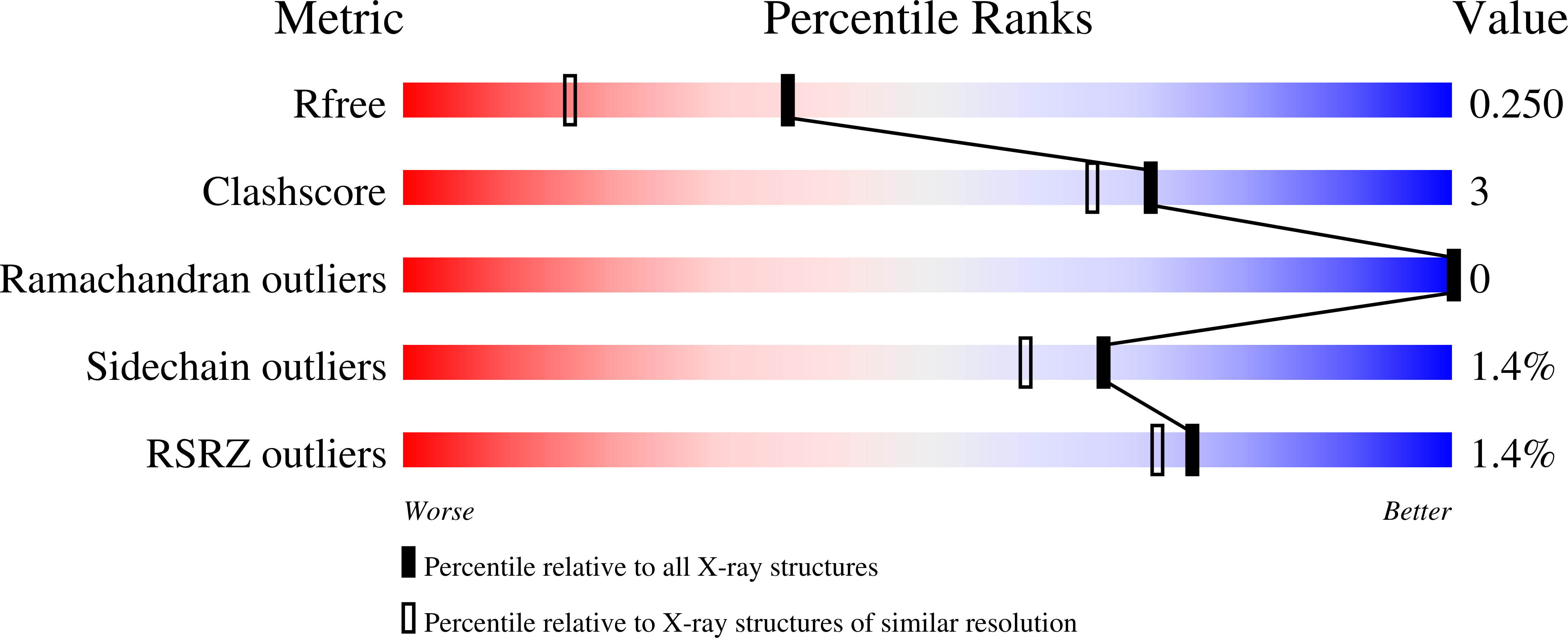
3GJ1 image
Deposition Date 2009-03-07
Release Date 2009-03-24
Last Version Date 2024-10-16
Entry Detail
PDB ID:
3GJ1
Title:
Non photoactivated state of PA-GFP
Biological Source:
Source Organism:
Host Organism:
Method Details:
Experimental Method:
Resolution:
1.80 Å
R-Value Free:
0.25
R-Value Work:
0.20
R-Value Observed:
0.21
Space Group:
P 21 21 21
Macromolecular Entities
Polymer Type:polypeptide(L)
Molecule:Green fluorescent protein
Gene (Uniprot):GFP
Mutations:Q80R, F99S, M153T, V163A, T203H
Chain IDs:A, B, C, D
Chain Length:231
Number of Molecules:4
Biological Source:Aequorea victoria
Modified Residue
Compound ID Chain ID Parent Comp ID Details 2D Image
CRO A GLY ?
Primary Citation
Structure and mechanism of the photoactivatable green fluorescent protein.
J.Am.Chem.Soc. 131 4176 4177 (2009)
PMID: 19278226 DOI: 10.1021/ja808851n

Abstact

Crystal structures of the photoactivatable green fluorescent protein T203H variant (PA-GFP) have been solved in the native and photoactivated states, which under 488 nm illumination are dark and brightly fluorescent, respectively. We demonstrate that photoactivation of PA-GFP is the result of a UV-induced decarboxylation of the Glu222 side chain that shifts the chromophore equilibrium to the anionic form. Coupled with the T203H mutation, which stabilizes the native PA-GFP neutral chromophore, Glu222 decarboxylation yields a 100-fold contrast enhancement relative to wild-type GFP (WT). Additionally, the structures provide insights into the spectroscopic differences between WT and PA-GFP steady-state fluorescence maxima and excited-state proton transfer dynamics.

Legend

Protein

Chemical

Disease

Primary Citation of related structures