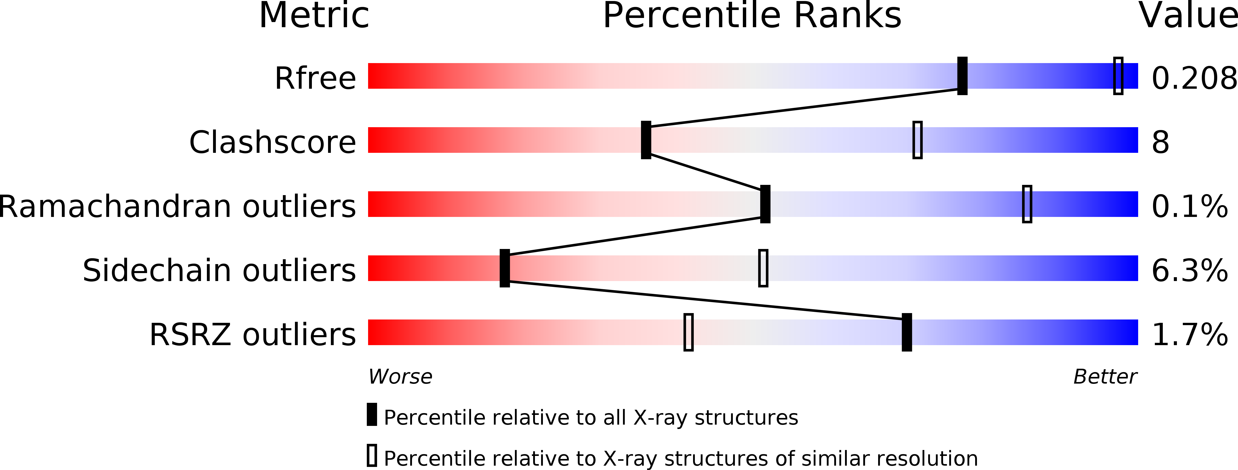
3GIT image
Deposition Date 2009-03-06
Release Date 2009-10-06
Last Version Date 2023-11-01
Entry Detail
PDB ID:
3GIT
Keywords:
Title:
Crystal structure of a truncated acetyl-CoA synthase
Biological Source:
Source Organism:
Method Details:
Experimental Method:
Resolution:
3.00 Å
R-Value Free:
0.20
R-Value Work:
0.17
R-Value Observed:
0.17
Space Group:
P 31 2 1
Macromolecular Entities
Polymer Type:polypeptide(L)
Molecule:Carbon monoxide dehydrogenase/acetyl-CoA synthase subunit alpha
Chain IDs:A, B, C, D, E, F
Chain Length:427
Number of Molecules:6
Biological Source:Moorella thermoacetica
Primary Citation
Novel domain arrangement in the crystal structure of a truncated acetyl-CoA synthase from Moorella thermoacetica
Biochemistry 48 7916 7926 (2009)
PMID: 19650626 DOI: 10.1021/bi9003952

Abstact

Ni-dependent acetyl-CoA synthase (ACS) and CO dehydrogenase (CODH) constitute the central enzyme complex of the Wood-Ljungdahl pathway of acetyl-CoA formation. The crystal structure of a recombinant bacterial ACS lacking the N-terminal domain that interacts with CODH shows a large reorganization of the remaining two globular domains, producing a narrow cleft of suitable size, shape, and nature to bind CoA. Sequence comparisons with homologous archaeal enzymes that naturally lack the N-terminal domain show that many amino acids lining this cleft are conserved. Besides the typical [4Fe-4S] center, the A-cluster contains only one proximal metal ion that, according to anomalous scattering data, is most likely Cu or Zn. Incorporation of a functional Ni(2)Fe(4)S(4) A-cluster would require only minor structural rearrangements. Using available structures, a plausible model of the interaction between CODH and the smaller ACS in archaeal multienzyme complexes is presented, along with a discussion of evolutionary relationships of the archaeal and bacterial enzymes.

Legend

Protein

Chemical

Disease

Primary Citation of related structures