Deposition Date
2009-03-05
Release Date
2009-06-02
Last Version Date
2024-11-27
Entry Detail
PDB ID:
3GIC
Keywords:
Title:
Structure of thrombin mutant delta(146-149e) in the free form
Biological Source:
Source Organism(s):
Homo sapiens (Taxon ID: 9606)
Expression System(s):
Method Details:
Experimental Method:
Resolution:
1.55 Å
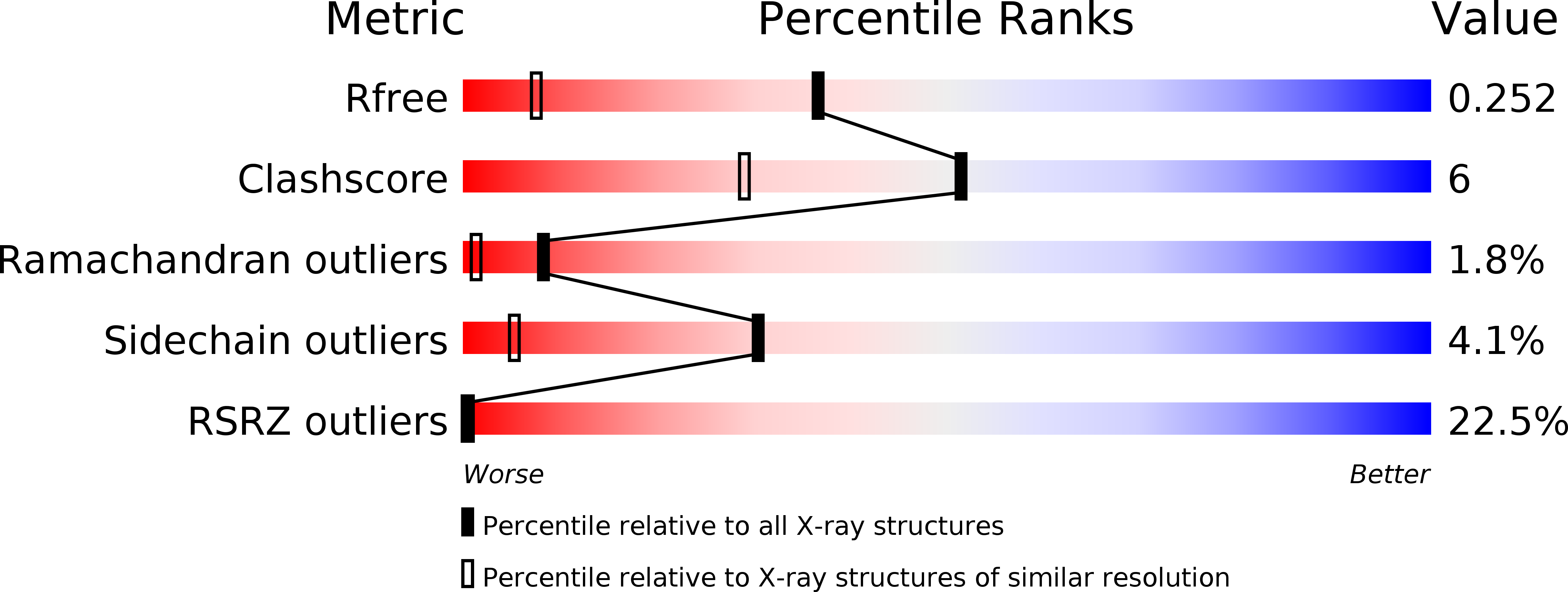
R-Value Free:
0.22
R-Value Work:
0.18
R-Value Observed:
0.18
Space Group:
P 43