3GGU image
Entry Detail
PDB ID:
3GGU
Keywords:
Title:
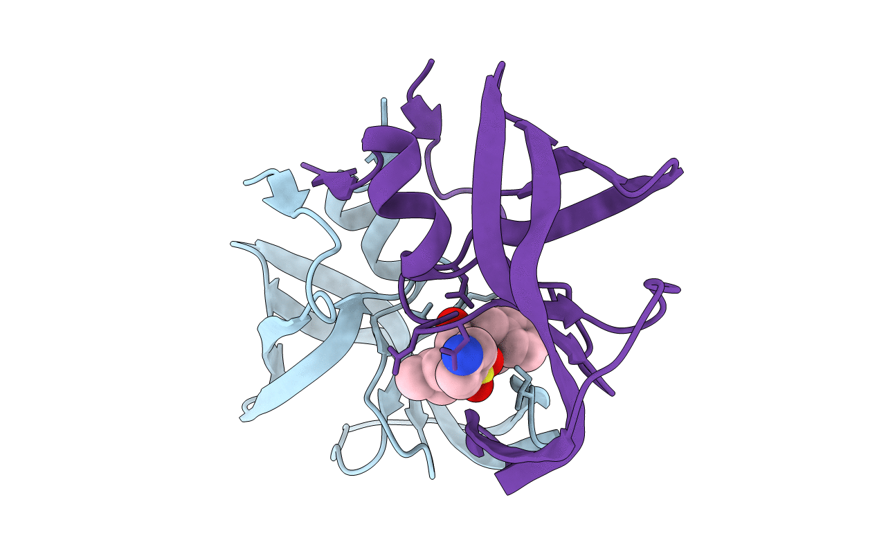
HIV PR drug resistant patient's variant in complex with darunavir
Biological Source:
PDB Version:
Deposition Date:
2009-03-02
Release Date:
2009-08-11
Method Details:
Experimental Method:
Resolution:
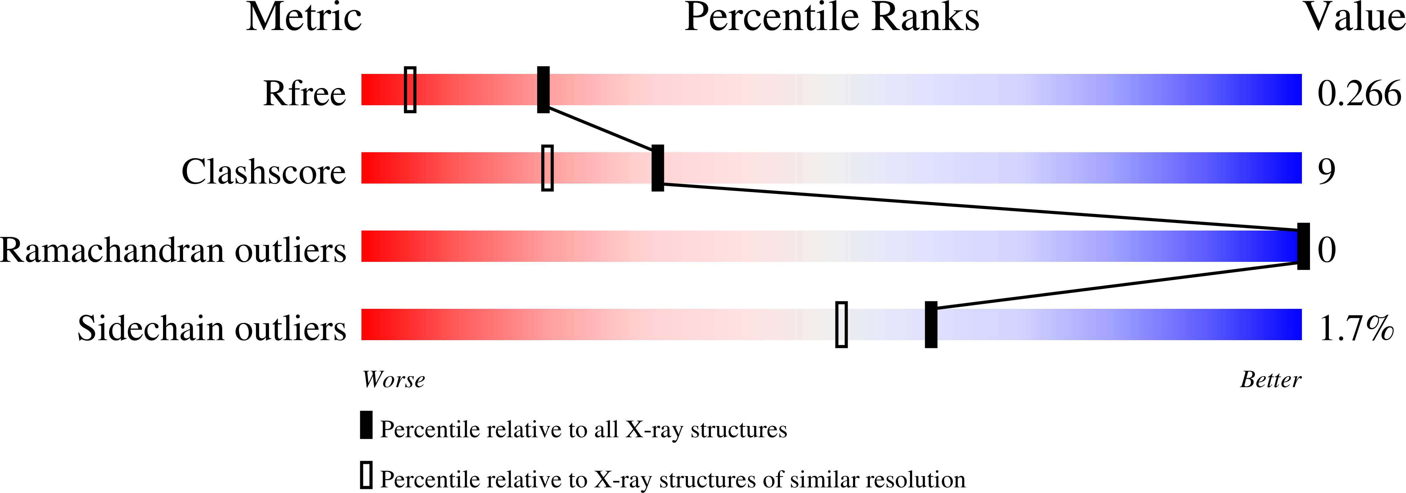
1.80 Å
R-Value Free:
0.24
R-Value Work:
0.20
R-Value Observed:
0.20
Space Group:
P 61
Macromolecular Entities
Polymer Type:polypeptide(L)
Description:Protease
Mutations:L10I, I13V, G16E, L33F, M36L, N37T, P39S, K45R, M46L, I54V, K55R, I62V, L63P, A71V, G73D, V82T, I84V, L89V, L90M, I93L
Chain IDs:A, B
Chain Length:99
Number of Molecules:2
Biological Source:Human immunodeficiency virus type 1 (BRU ISOLATE)
Ligand Molecules
Primary Citation
Molecular characterization of clinical isolates of human immunodeficiency virus resistant to the protease inhibitor darunavir.
J.Virol. 83 8810 8818 (2009)
PMID: 19535439 DOI: 10.1128/JVI.00451-09

Abstact

Darunavir is the most recently approved human immunodeficiency virus (HIV) protease (PR) inhibitor (PI) and is active against many HIV type 1 PR variants resistant to earlier-generation PIs. Darunavir shows a high genetic barrier to resistance development, and virus strains with lower sensitivity to darunavir have a higher number of PI resistance-associated mutations than viruses resistant to other PIs. In this work, we have enzymologically and structurally characterized a number of highly mutated clinically derived PRs with high levels of phenotypic resistance to darunavir. With 18 to 21 amino acid residue changes, the PR variants studied in this work are the most highly mutated HIV PR species ever studied by means of enzyme kinetics and X-ray crystallography. The recombinant proteins showed major defects in substrate binding, while the substrate turnover was less affected. Remarkably, the overall catalytic efficiency of the recombinant PRs (5% that of the wild-type enzyme) is still sufficient to support polyprotein processing and particle maturation in the corresponding viruses. The X-ray structures of drug-resistant PRs complexed with darunavir suggest that the impaired inhibitor binding could be explained by change in the PR-inhibitor hydrogen bond pattern in the P2' binding pocket due to a substantial shift of the aminophenyl moiety of the inhibitor. Recombinant virus phenotypic characterization, enzyme kinetics, and X-ray structural analysis thus help to explain darunavir resistance development in HIV-positive patients.

Legend

Protein

Chemical

Disease

Primary Citation of related structures