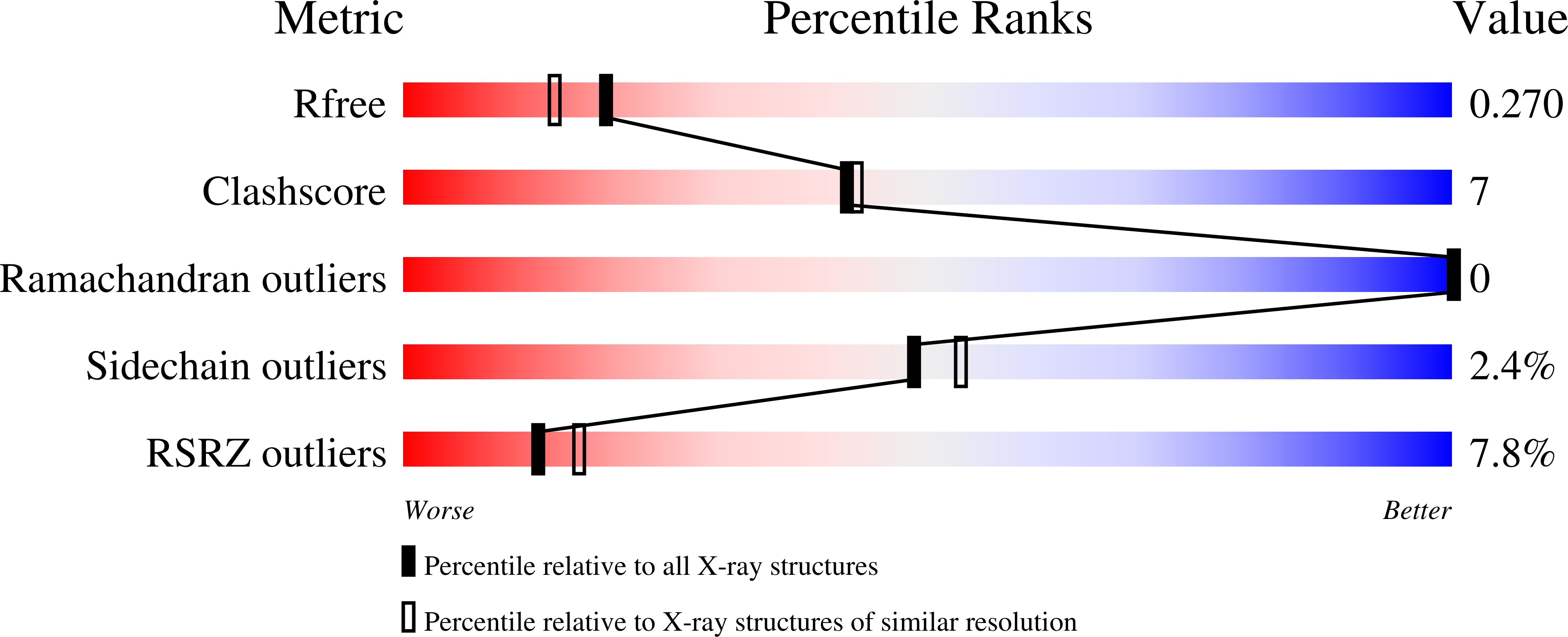
3GFM image
Deposition Date 2009-02-27
Release Date 2009-08-25
Last Version Date 2023-11-01
Entry Detail
PDB ID:
3GFM
Keywords:
Title:
Crystal structure of the ST1710 mutant (K91A) protein
Biological Source:
Source Organism:
Host Organism:
Method Details:
Experimental Method:
Resolution:
2.10 Å
R-Value Free:
0.27
R-Value Work:
0.21
R-Value Observed:
0.21
Space Group:
P 41 21 2
Macromolecular Entities
Polymer Type:polypeptide(L)
Molecule:146aa long hypothetical transcriptional regulator
Gene (Uniprot):ST1710
Mutations:K91A
Chain IDs:A
Chain Length:146
Number of Molecules:1
Biological Source:Sulfolobus tokodaii
Ligand Molecules
Primary Citation
ST1710-DNA complex crystal structure reveals the DNA binding mechanism of the MarR family of regulators.
Nucleic Acids Res. 37 4723 4735 (2009)
PMID: 19509310 DOI: 10.1093/nar/gkp496

Abstact

ST1710, a member of the multiple antibiotic resistance regulator (MarR) family of regulatory proteins in bacteria and archaea, plays important roles in development of antibiotic resistance, a global health problem. Here, we present the crystal structure of ST1710 from Sulfolobus tokodaii strain 7 complexed with salicylate, a well-known inhibitor of MarR proteins and the ST1710 complex with its promoter DNA, refined to 1.8 and 2.10 A resolutions, respectively. The ST1710-DNA complex shares the topology of apo-ST1710 and MarR proteins, with each subunit containing a winged helix-turn-helix (wHtH) DNA binding motif. Significantly large conformational changes occurred upon DNA binding and in each of the dimeric monomers in the asymmetric unit of the ST1710-DNA complex. Conserved wHtH loop residues interacting with the bound DNA and mutagenic analysis indicated that R89, R90 and K91 were important for DNA recognition. Significantly, the bound DNA exhibited a new binding mechanism.

Legend

Protein

Chemical

Disease

Primary Citation of related structures