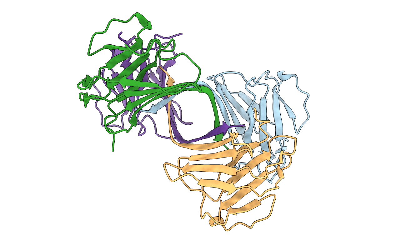
3GEF image
Deposition Date 2009-02-25
Release Date 2009-08-04
Last Version Date 2024-11-27
Entry Detail
PDB ID:
3GEF
Title:
Crystal structure of the R482W mutant of lamin A/C
Biological Source:
Source Organism:
Homo sapiens (Taxon ID: 9606)
Host Organism:
Method Details:
Experimental Method:
Resolution:
1.50 Å
R-Value Free:
0.25
R-Value Work:
0.21
Space Group:
P 21 21 21
Macromolecular Entities
Polymer Type:polypeptide(L)
Molecule:Lamin-A/C
Gene (Uniprot):LMNA
Mutations:R482W
Chain IDs:A, B, C, D
Chain Length:118
Number of Molecules:4
Biological Source:Homo sapiens
Modified Residue
Compound ID Chain ID Parent Comp ID Details 2D Image
CSD A CYS 3-SULFINOALANINE
Primary Citation
Structure of the lamin A/C R482W mutant responsible for dominant familial partial lipodystrophy (FPLD).
Acta Crystallogr.,Sect.F 65 665 670 (2009)
PMID: 19574635 DOI: 10.1107/S1744309109020302

Abstact

Proteins of the A-type lamin family, which consists of two members, lamin A and lamin C, are the major components of a thin proteinaceous filamentous meshwork, the lamina, that underlies the inner nuclear membrane. A-type lamins have recently become the focus of extensive functional studies as a consequence of the linking of at least eight congenital diseases to mutations in the lamin A/C gene (LMNA). This spectrum of pathologies, which mostly manifest themselves as dominant traits, includes muscle dystrophies, dilated cardiomyopathies, the premature aging syndrome Hutchinson-Guilford progeria and familial partial lipodystrophy (FPLD). The crystal structure of the lamin A/C mutant R482W, a variant that causes FPLD, has been determined at 1.5 A resolution. A completely novel aggregation state of the C-terminal globular domain and the position of the mutated amino-acid residue suggest means by which the mutation may affect lamin A/C-protein and protein-DNA interactions.

Legend

Protein

Chemical

Disease

Primary Citation of related structures