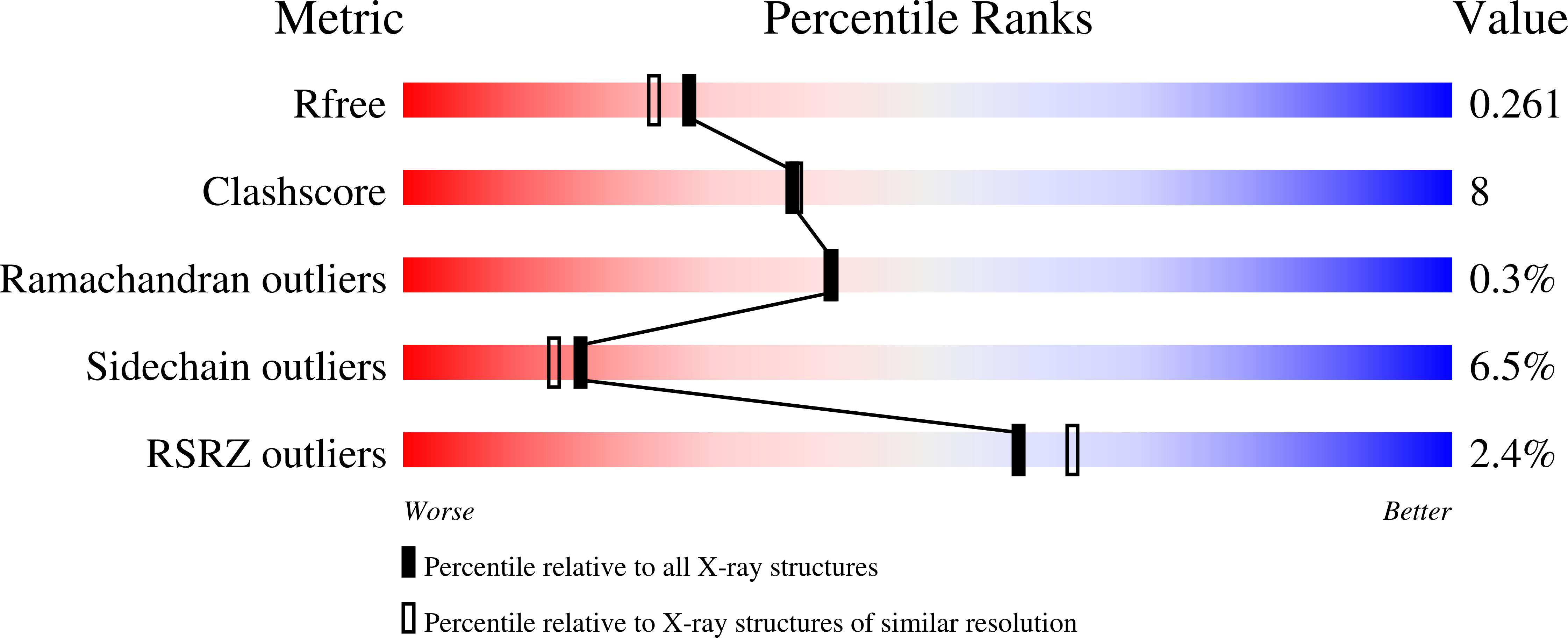
3GCS image
Deposition Date 2009-02-22
Release Date 2009-06-09
Last Version Date 2023-09-06
Entry Detail
PDB ID:
3GCS
Keywords:
Title:
Human P38 MAP kinase in complex with Sorafenib
Biological Source:
Source Organism:
Homo sapiens (Taxon ID: 9606)
Host Organism:
Method Details:
Experimental Method:
Resolution:
2.10 Å
R-Value Free:
0.26
R-Value Work:
0.20
R-Value Observed:
0.20
Space Group:
P 21 21 21
Macromolecular Entities
Polymer Type:polypeptide(L)
Molecule:Mitogen-activated protein kinase 14
Gene (Uniprot):MAPK14
Mutations:C119S, C162S, A172C, F327L
Chain IDs:A
Chain Length:360
Number of Molecules:1
Biological Source:Homo sapiens
Primary Citation
Development of a fluorescent-tagged kinase assay system for the detection and characterization of allosteric kinase inhibitors.
J.Am.Chem.Soc. 131 13286 13296 (2009)
PMID: 19572644 DOI: 10.1021/ja902010p

Abstact

Kinase disregulation disrupts the intricate network of intracellular signaling pathways and contributes to the onset of diseases such as cancer. Although several kinase inhibitors are on the market, inhibitor selectivity and drug resistance mutations persist as fundamental challenges in the development of effective long-term treatments. Chemical entities binding to less conserved allosteric sites would be expected to offer new opportunities for scaffold development. Because no high-throughput method was previously available, we developed a fluorescence-based kinase binding assay for identifying and characterizing ligands which stabilize the inactive kinase conformation. Here, we present a description of the development and validation of this assay using the serine/threonine kinase p38alpha. By covalently attaching fluorophores to the activation loop of the kinase, we were able to detect conformational changes and measure the K(d), k(on), and k(off) associated with the binding and dissociation of ligands to the allosteric pocket. We report the SAR of a synthesized focused library of pyrazolourea derivatives, a scaffold known to bind with high affinity to the allosteric pocket of p38alpha. Additionally, we used protein X-ray crystallography together with our assay to examine the binding and dissociation kinetics to characterize potent quinazoline- and quinoline-based type II inhibitors, which also utilize this binding pocket in p38alpha. Last, we identified the b-Raf inhibitor sorafenib as a potent low nanomolar inhibitor of p38alpha and used protein X-ray crystallography to confirm a unique binding mode to the inactive kinase conformation.

Legend

Protein

Chemical

Disease

Primary Citation of related structures