
Deposition Date
2009-02-21
Release Date
2009-07-21
Last Version Date
2023-09-06
Entry Detail
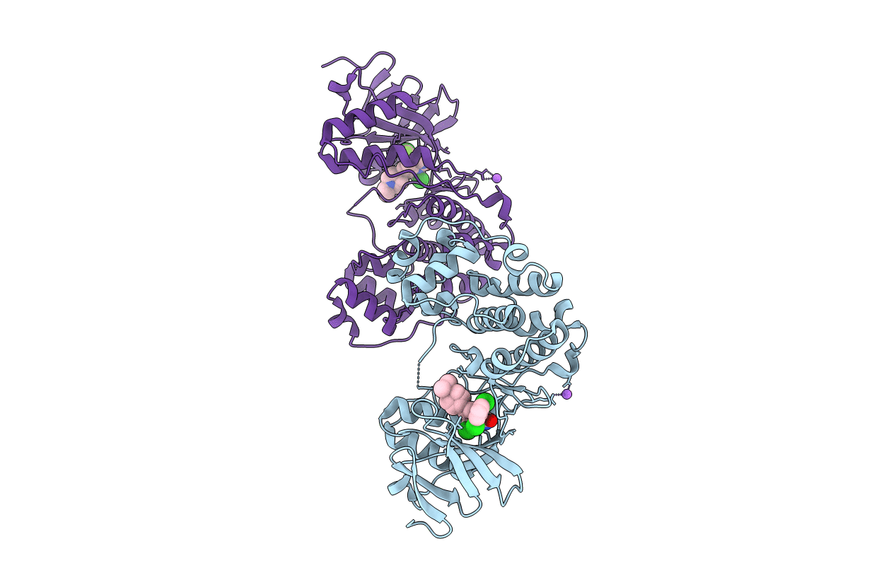
PDB ID:
3GC9
Keywords:
Title:
The structure of p38beta C119S, C162S in complex with a dihydroquinazolinone inhibitor
Biological Source:
Source Organism(s):
Homo sapiens (Taxon ID: 9606)
Expression System(s):
Method Details:
Experimental Method:
Resolution:
2.05 Å
R-Value Free:
0.27
R-Value Work:
0.22
R-Value Observed:
0.22
Space Group:
P 1 21 1
