Deposition Date
2009-02-16
Release Date
2009-04-14
Last Version Date
2023-11-01
Entry Detail
PDB ID:
3GA5
Keywords:
Title:
X-ray structure of glucose/galactose receptor from Salmonella typhimurium in complex with (2R)-glyceryl-beta-D-galactopyranoside
Biological Source:
Source Organism(s):
Salmonella typhimurium (Taxon ID: 602)
Expression System(s):
Method Details:
Experimental Method:
Resolution:
1.87 Å
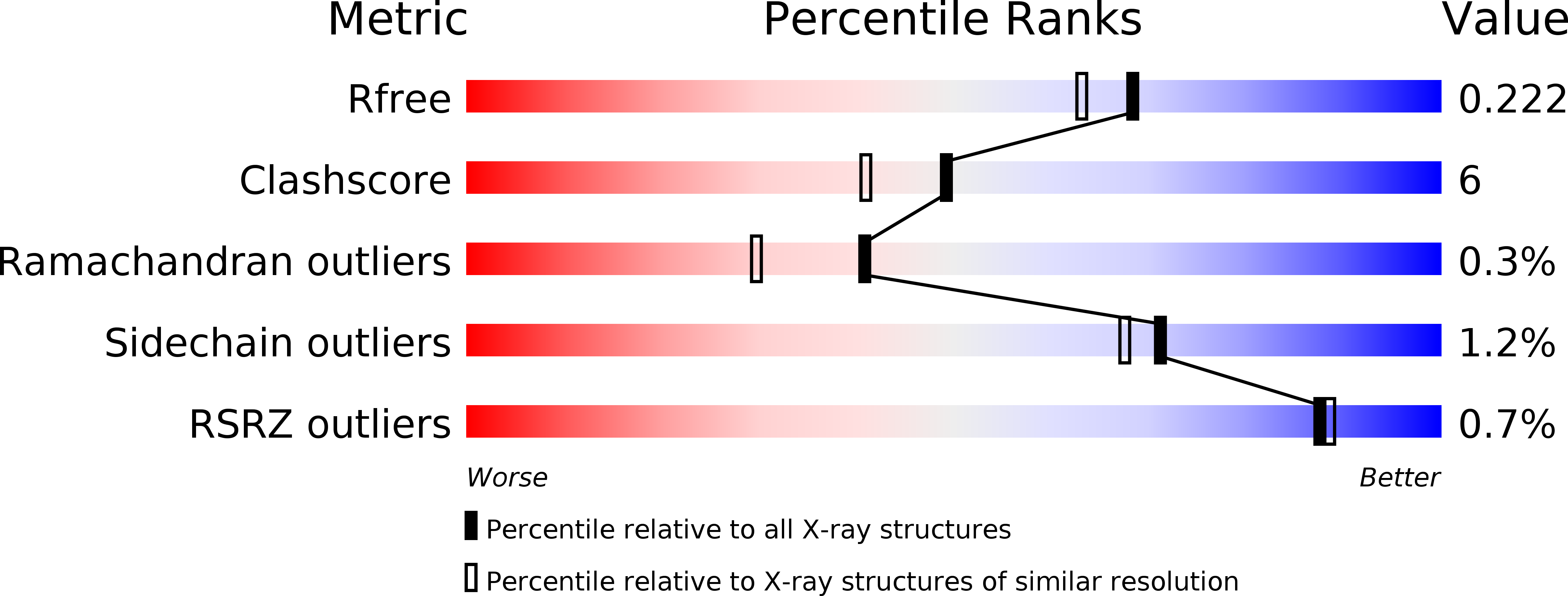
R-Value Free:
0.22
R-Value Work:
0.17
R-Value Observed:
0.17
Space Group:
P 21 21 21