3G7F image
Deposition Date 2009-02-09
Release Date 2009-09-22
Last Version Date 2024-11-20
Entry Detail
PDB ID:
3G7F
Keywords:
Title:
Crystal structure of Blastochloris viridis heterodimer mutant reaction center
Biological Source:
Source Organism(s):
Method Details:
Experimental Method:
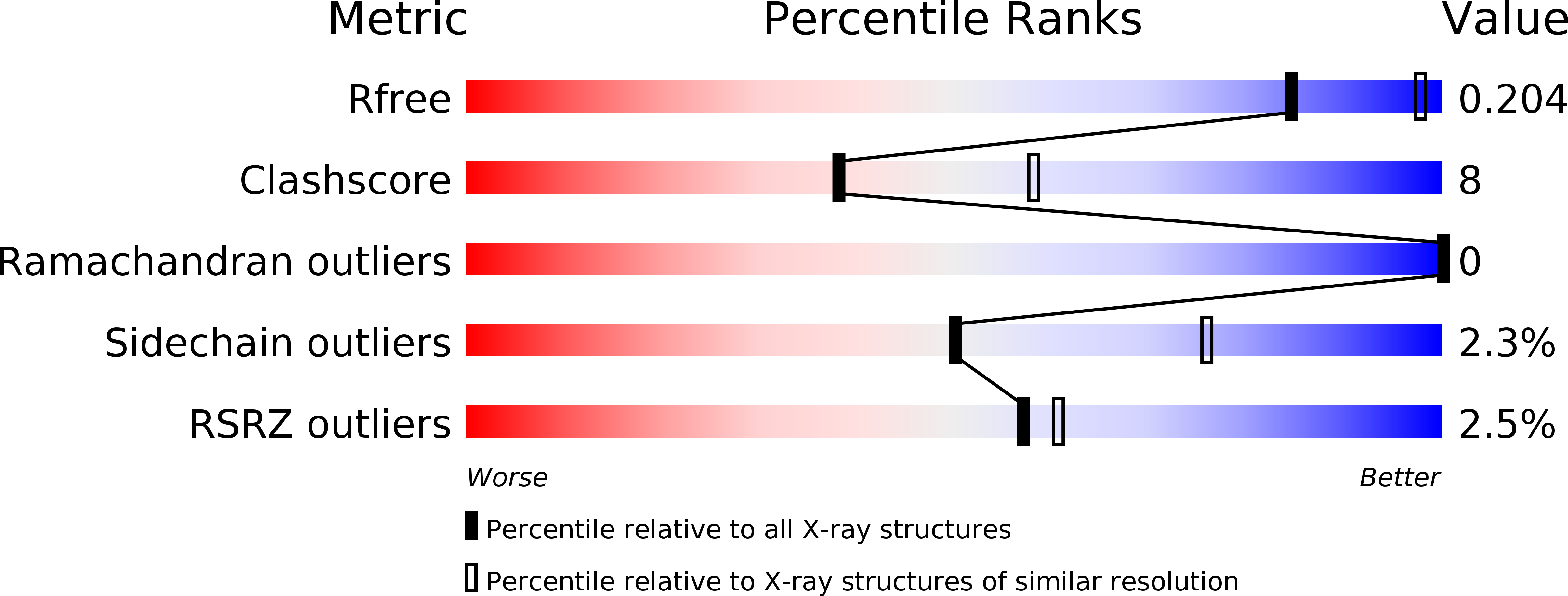
Resolution:
2.50 Å
R-Value Free:
0.20
R-Value Work:
0.17
R-Value Observed:
0.17
Space Group:
P 43 21 2
Macromolecular Entities
Structures with similar UniProt ID
Protein Blast
Polymer Type:polypeptide(L)
Molecule:Photosynthetic reaction center cytochrome c subunit
Gene (Uniprot):pufC
Chain IDs:A (auth: C)
Chain Length:336
Number of Molecules:1
Biological Source:Blastochloris viridis
Structures with similar UniProt ID
Protein Blast
Polymer Type:polypeptide(L)
Molecule:Photosynthetic reaction center H subunit
Gene (Uniprot):puhA
Chain IDs:B (auth: H)
Chain Length:258
Number of Molecules:1
Biological Source:Blastochloris viridis
Structures with similar UniProt ID
Protein Blast
Polymer Type:polypeptide(L)
Molecule:Photosynthetic reaction center L subunit
Gene (Uniprot):pufL
Chain IDs:C (auth: L)
Chain Length:273
Number of Molecules:1
Biological Source:Blastochloris viridis
Structures with similar UniProt ID
Protein Blast
Polymer Type:polypeptide(L)
Molecule:Photosynthetic reaction center M subunit
Gene (Uniprot):pufM
Chain IDs:D (auth: M)
Chain Length:323
Number of Molecules:1
Biological Source:Blastochloris viridis
Modified Residue
Compound ID Chain ID Parent Comp ID Details 2D Image
FME B MET N-FORMYLMETHIONINE
Primary Citation
Structural and spectropotentiometric analysis of Blastochloris viridis heterodimer mutant reaction center
Biochim.Biophys.Acta 1788 1822 1831 (2009)
PMID: 19539602 DOI: 10.1016/j.bbamem.2009.06.006

Abstact

Heterodimer mutant reaction centers (RCs) of Blastochloris viridis were crystallized using microfluidic technology. In this mutant, a leucine residue replaced the histidine residue which had acted as a fifth ligand to the bacteriochlorophyll (BChl) of the primary electron donor dimer M site (HisM200). With the loss of the histidine-coordinated Mg, one bacteriochlorophyll of the special pair was converted into a bacteriopheophytin (BPhe), and the primary donor became a heterodimer supermolecule. The crystals had dimensions 400 x 100 x 100 microm, belonged to space group P4(3)2(1)2, and were isomorphous to the ones reported earlier for the wild type (WT) strain. The structure was solved to a 2.5 A resolution limit. Electron-density maps confirmed the replacement of the histidine residue and the absence of Mg. Structural changes in the heterodimer mutant RC relative to the WT included the absence of the water molecule that is typically positioned between the M side of the primary donor and the accessory BChl, a slight shift in the position of amino acids surrounding the site of the mutation, and the rotation of the M194 phenylalanine. The cytochrome subunit was anchored similarly as in the WT and had no detectable changes in its overall position. The highly conserved tyrosine L162, located between the primary donor and the highest potential heme C(380), revealed only a minor deviation of its hydroxyl group. Concomitantly to modification of the BChl molecule, the redox potential of the heterodimer primary donor increased relative to that of the WT organism (772 mV vs. 517 mV). The availability of this heterodimer mutant and its crystal structure provides opportunities for investigating changes in light-induced electron transfer that reflect differences in redox cascades.

Legend

Protein

Chemical

Disease

Primary Citation of related structures
Feedback Form
Name
Email
Institute
Feedback