3G5I image
Deposition Date 2009-02-05
Release Date 2010-02-09
Last Version Date 2023-11-01
Entry Detail
PDB ID:
3G5I
Keywords:
Title:
Crystal Structure of the E.coli RihA pyrimidine nucleosidase bound to a iminoribitol-based inhibitor
Biological Source:
Source Organism:
Host Organism:
Method Details:
Experimental Method:
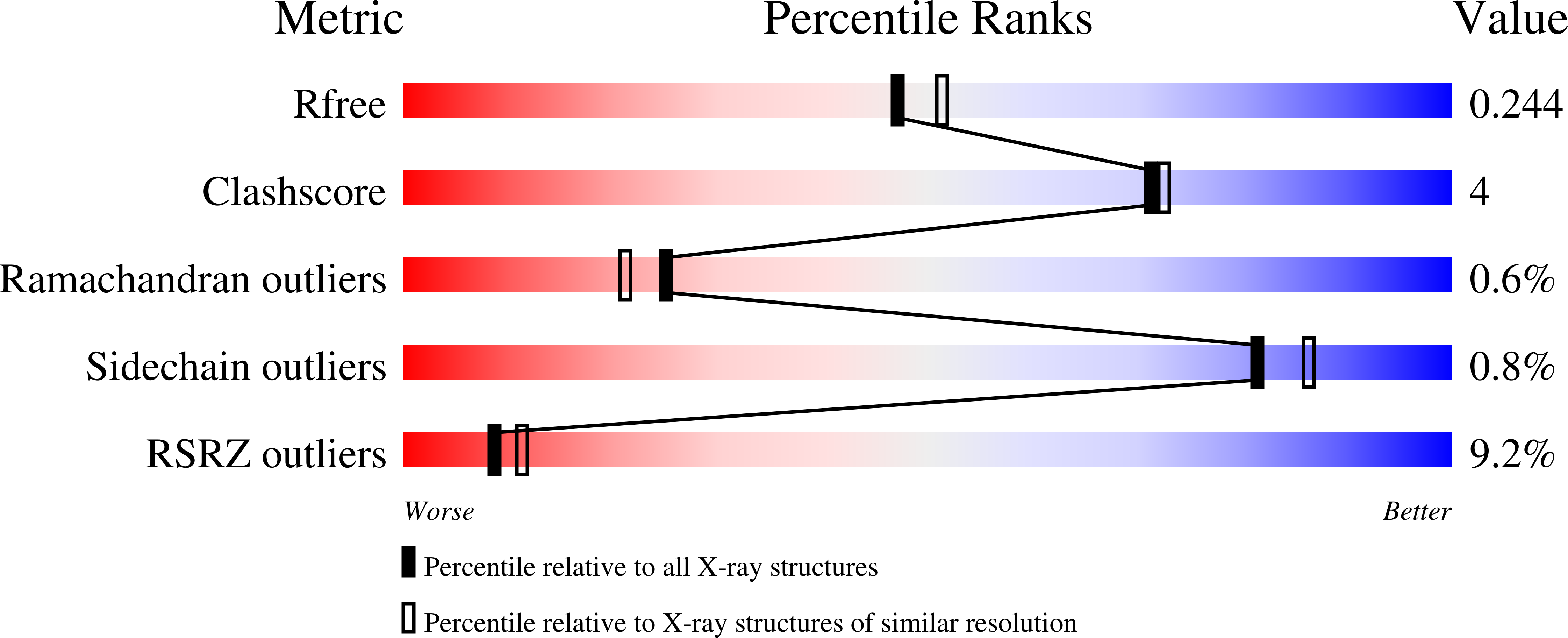
Resolution:
2.10 Å
R-Value Free:
0.24
R-Value Work:
0.20
R-Value Observed:
0.20
Space Group:
P 1 21 1
Macromolecular Entities
Structures with similar UniProt ID
Protein Blast
Polymer Type:polypeptide(L)
Molecule:Pyrimidine-specific ribonucleoside hydrolase rihA
Gene (Uniprot):rihA
Chain IDs:A, B, C, D
Chain Length:312
Number of Molecules:4
Biological Source:Escherichia coli
Primary Citation
Active site plasticity revealed from the structure of the enterobacterial N-ribohydrolase RihA bound to a competitive inhibitor.
Bmc Struct.Biol. 10 14 14 (2010)
PMID: 20529317 DOI: 10.1186/1472-6807-10-14

Abstact

BACKGROUND Pyrimidine-preferring N-ribohydrolases (CU-NHs) are a class of Ca2+-dependent enzymes that catalyze the hydrolytic cleavage of the N-glycosidic bond in pyrimidine nucleosides. With the exception of few selected organisms, their physiological relevance in prokaryotes and eukaryotes is yet under investigation. RESULTS Here, we report the first crystal structure of a CU-NH bound to a competitive inhibitor, the complex between the Escherichia coli enzyme RihA bound to 3, 4-diaminophenyl-iminoribitol (DAPIR) to a resolution of 2.1 A. The ligand can bind at the active site in two distinct orientations, and the stabilization of two flexible active site regions is pivotal to establish the interactions required for substrate discrimination and catalysis. CONCLUSIONS A comparison with the product-bound RihA structure allows a rationalization of the structural rearrangements required for an enzymatic catalytic cycle, highlighting a substrate-assisted cooperative motion, and suggesting a yet overlooked role of the conserved His82 residue in modulating product release. Differences in the structural features of the active sites in the two homologous CU-NHs RihA and RihB from E. coli provide a rationale for their fine differences in substrate specificity. These new findings hint at a possible role of CU-NHs in the breakdown of modified nucleosides derived from RNA molecules.

Legend

Protein

Chemical

Disease

Primary Citation of related structures