3G4V image
Entry Detail
PDB ID:
3G4V
Keywords:
Title:
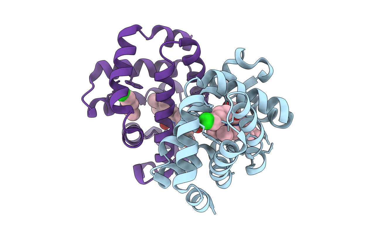
Ligand migration and cavities within scapharca dimeric hemoglobin: wild type with co bound to heme and chloropentane bound to the XE4 cavity
Biological Source:
Source Organism:
Host Organism:
PDB Version:
Deposition Date:
2009-02-04
Release Date:
2009-12-01
Method Details:
Experimental Method:
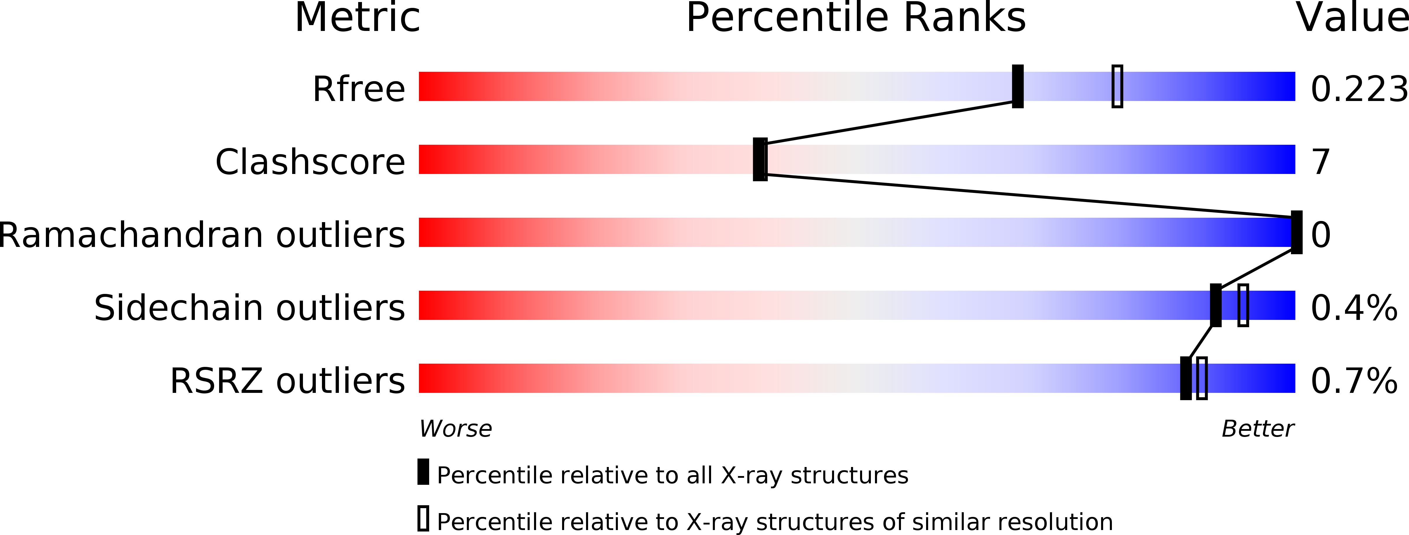
Resolution:
2.10 Å
R-Value Free:
0.23
R-Value Work:
0.19
R-Value Observed:
0.19
Space Group:
C 1 2 1
Macromolecular Entities
Polymer Type:polypeptide(L)
Description:GLOBIN-1
Chain IDs:A, B
Chain Length:146
Number of Molecules:2
Biological Source:Scapharca inaequivalvis
Primary Citation
Ligand migration and cavities within Scapharca Dimeric HbI: studies by time-resolved crystallo-graphy, Xe binding, and computational analysis.
Structure 17 1494 1504 (2009)
PMID: 19913484 DOI: 10.1016/j.str.2009.09.004

Abstact

As in many other hemoglobins, no direct route for migration of ligands between solvent and active site is evident from crystal structures of Scapharca inaequivalvis dimeric HbI. Xenon (Xe) and organic halide binding experiments, along with computational analysis presented here, reveal protein cavities as potential ligand migration routes. Time-resolved crystallographic experiments show that photodissociated carbon monoxide (CO) docks within 5 ns at the distal pocket B site and at more remote Xe4 and Xe2 cavities. CO rebinding is not affected by the presence of dichloroethane within the major Xe4 protein cavity, demonstrating that this cavity is not on the major exit pathway. The crystal lattice has a substantial influence on ligand migration, suggesting that significant conformational rearrangements may be required for ligand exit. Taken together, these results are consistent with a distal histidine gate as one important ligand entry and exit route, despite its participation in the dimeric interface.

Legend

Protein

Chemical

Disease

Primary Citation of related structures