Deposition Date
2009-02-03
Release Date
2010-02-02
Last Version Date
2023-09-06
Entry Detail
PDB ID:
3G4E
Keywords:
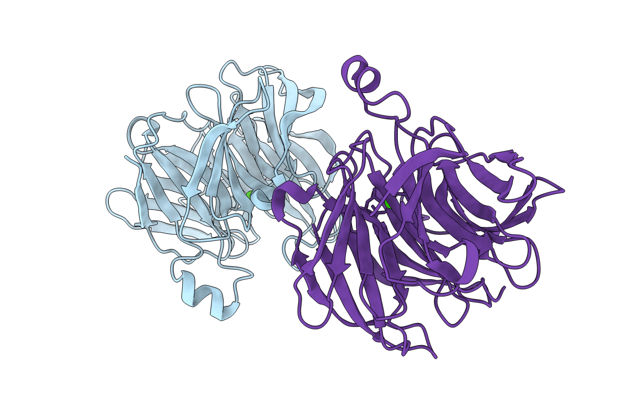
Title:
Crystal structure of human senescence marker protein-30(SMP30)(Calcium bound)
Biological Source:
Source Organism(s):
Homo sapiens (Taxon ID: 9606)
Expression System(s):
Method Details:
Experimental Method:
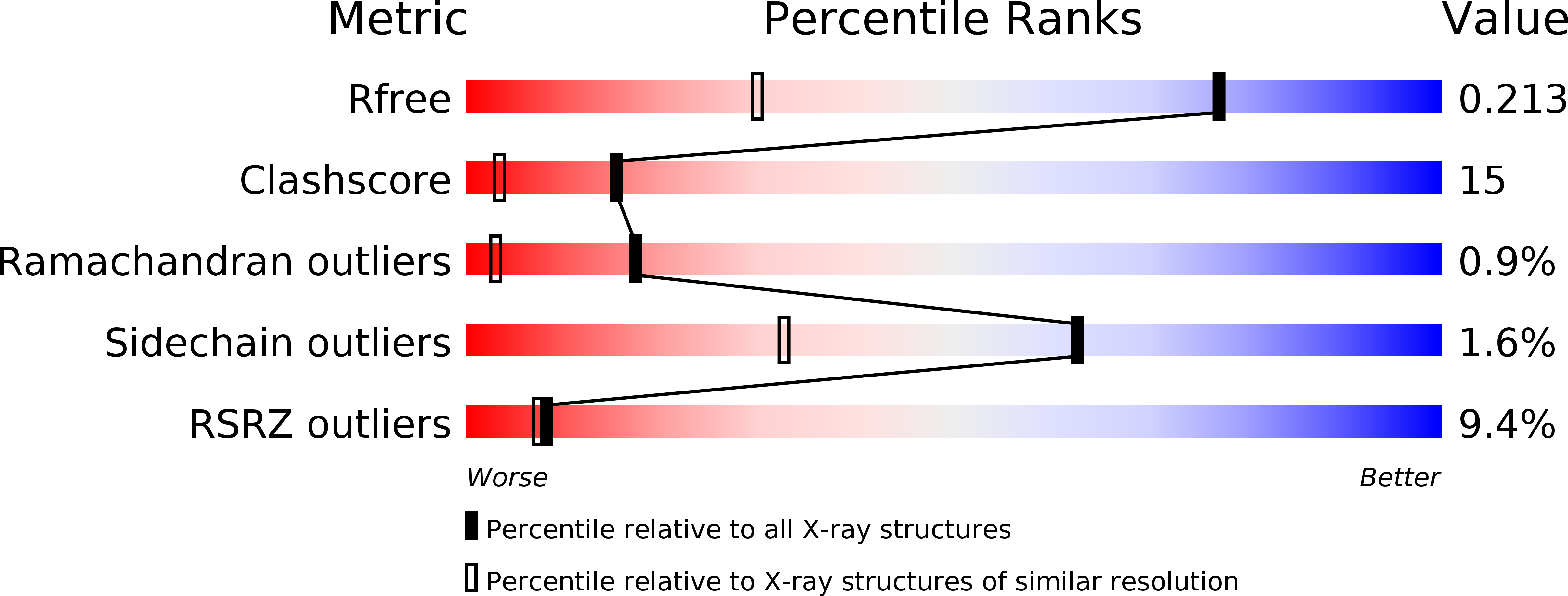
Resolution:
1.42 Å
R-Value Free:
0.19
R-Value Work:
0.12
R-Value Observed:
0.13
Space Group:
P 1 21 1