3G3V image
Deposition Date 2009-02-02
Release Date 2009-05-05
Last Version Date 2024-10-30
Entry Detail
PDB ID:
3G3V
Keywords:
Title:
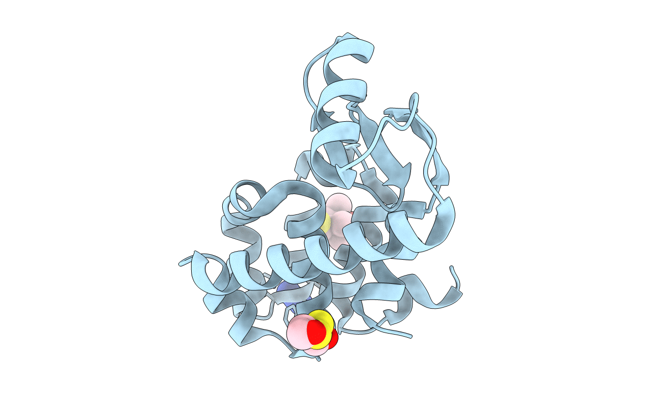
Crystal structure of spin labeled T4 Lysozyme (V131R1) at 291 K
Biological Source:
Source Organism:
Host Organism:
Method Details:
Experimental Method:
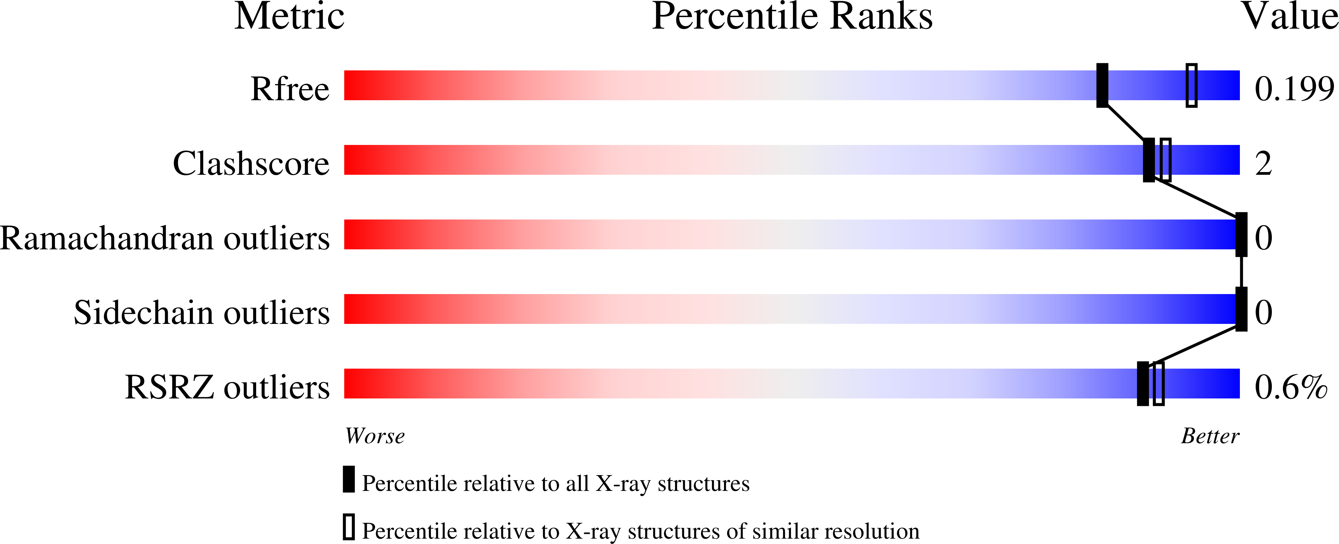
Resolution:
2.10 Å
R-Value Free:
0.19
R-Value Work:
0.14
R-Value Observed:
0.14
Space Group:
P 32 2 1
Macromolecular Entities
Structures with similar UniProt ID
Protein Blast
Polymer Type:polypeptide(L)
Molecule:Lysozyme
Gene (Uniprot):E
Mutagens:C54T, C97A, V131C
Chain IDs:A
Chain Length:164
Number of Molecules:1
Biological Source:Enterobacteria phage T4
Primary Citation
Structural origin of weakly ordered nitroxide motion in spin-labeled proteins.
Protein Sci. 18 893 908 (2009)
PMID: 19384990 DOI: 10.1002/pro.96

Abstact

A disulfide-linked nitroxide side chain (R1) used in site-directed spin labeling of proteins often exhibits an EPR spectrum characteristic of a weakly ordered z-axis anisotropic motion at topographically diverse surface sites, including those on helices, loops and edge strands of beta-sheets. To elucidate the origin of this motion, the first crystal structures of R1 that display simple z-axis anisotropic motion at solvent-exposed helical sites (131 and 151) and a loop site (82) in T4 lysozyme have been determined. Structures of 131R1 and 151R1 determined at cryogenic or ambient temperature reveal an intraresidue C(alpha)--H...S(delta) interaction that immobilizes the disulfide group, consistent with a model in which the internal motions of R1 are dominated by rotations about the two terminal bonds (Columbus, Kálai, Jeko, Hideg, and Hubbell, Biochemistry 2001;40:3828-3846). Remarkably, the 131R1 side chain populates two rotamers equally, but the EPR spectrum reflects a single dominant dynamic population, showing that the two rotamers have similar internal motion determined by the common disulfide-backbone interaction. The anisotropic motion for loop residue 82R1 is also accounted for by a common disulfide-backbone interaction, showing that the interaction does not require a specific secondary structure. If the above observations prove to be general, then significant variations in order and rate for R1 at noninteracting solvent-exposed helical and loop sites can be assigned to backbone motion because the internal motion is essentially constant.

Legend

Protein

Chemical

Disease

Primary Citation of related structures