3G05 image
Entry Detail
PDB ID:
3G05
Title:
Crystal structure of N-terminal domain (2-550) of E.coli MnmG
Biological Source:
Host Organism:
PDB Version:
Deposition Date:
2009-01-27
Release Date:
2009-10-20
Method Details:
Experimental Method:
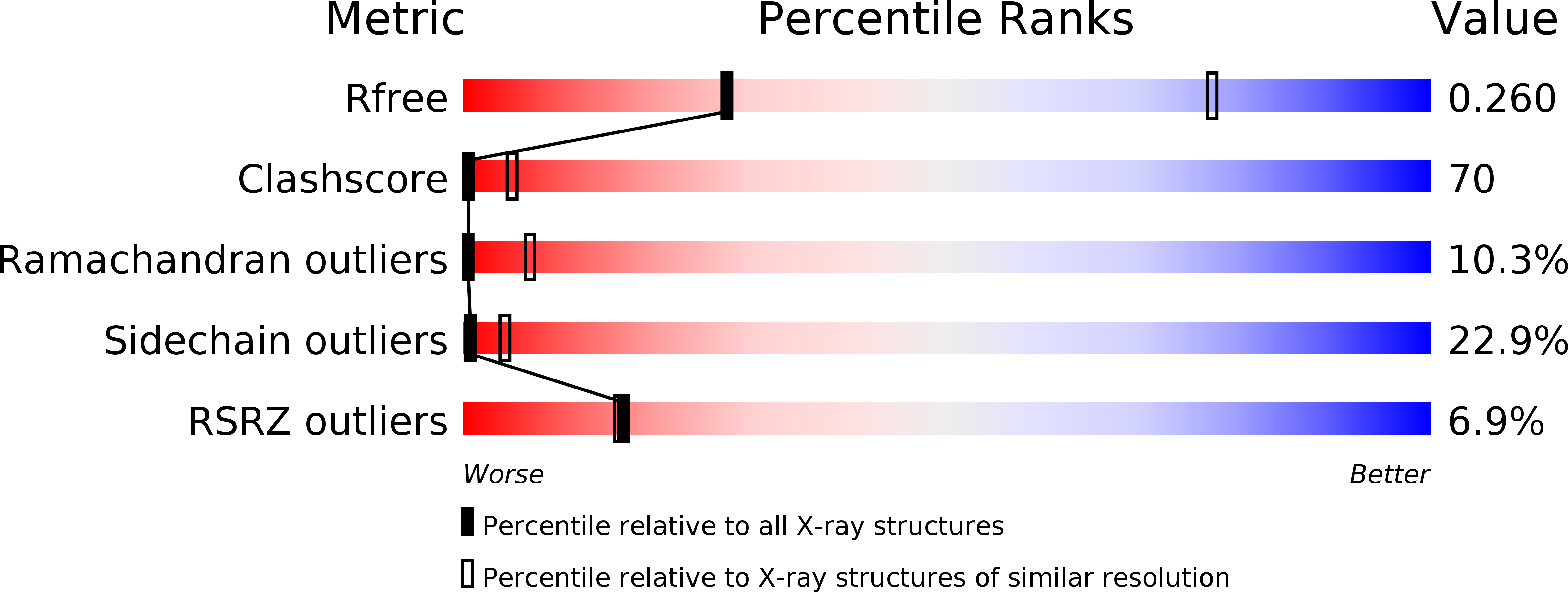
Resolution:
3.49 Å
R-Value Free:
0.26
R-Value Work:
0.22
R-Value Observed:
0.22
Space Group:
P 31 2 1
Macromolecular Entities
Polymer Type:polypeptide(L)
Description:tRNA uridine 5-carboxymethylaminomethyl modification enzyme mnmG
Chain IDs:A, B
Chain Length:576
Number of Molecules:2
Biological Source:Escherichia coli O157:H7 EDL933
Ligand Molecules
Primary Citation
Structure-function analysis of Escherichia coli MnmG (GidA), a highly conserved tRNA-modifying enzyme.
J.Bacteriol. 191 7614 7619 (2009)
PMID: 19801413 DOI: 10.1128/JB.00650-09

Abstact

The MnmE-MnmG complex is involved in tRNA modification. We have determined the crystal structure of Escherichia coli MnmG at 2.4-A resolution, mutated highly conserved residues with putative roles in flavin adenine dinucleotide (FAD) or tRNA binding and MnmE interaction, and analyzed the effects of these mutations in vivo and in vitro. Limited trypsinolysis of MnmG suggests significant conformational changes upon FAD binding.

Legend

Protein

Chemical

Disease

Primary Citation of related structures