3G03 image
Deposition Date 2009-01-27
Release Date 2009-04-14
Last Version Date 2023-11-01
Entry Detail
PDB ID:
3G03
Keywords:
Title:
Structure of human MDM2 in complex with high affinity peptide
Biological Source:
Source Organism:
Homo sapiens (Taxon ID: 9606)
Host Organism:
Method Details:
Experimental Method:
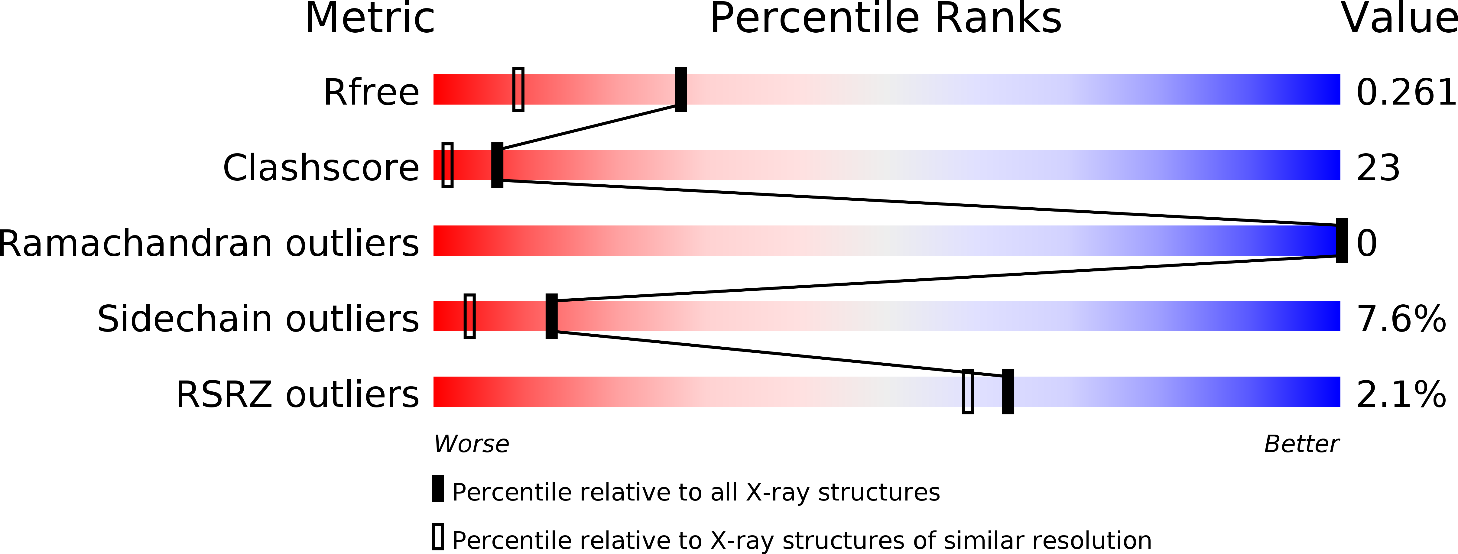
Resolution:
1.80 Å
R-Value Free:
0.26
R-Value Work:
0.21
R-Value Observed:
0.21
Space Group:
P 21 21 21
Macromolecular Entities
Structures with similar UniProt ID
Protein Blast
Polymer Type:polypeptide(L)
Molecule:E3 ubiquitin-protein ligase Mdm2
Gene (Uniprot):MDM2
Chain IDs:A, C
Chain Length:109
Number of Molecules:2
Biological Source:Homo sapiens
Polymer Type:polypeptide(L)
Molecule:High affinity synthetic peptide
Chain IDs:B, D
Chain Length:12
Number of Molecules:2
Biological Source:
Primary Citation
High affinity interaction of the p53 peptide-analogue with human Mdm2 and Mdmx.
Cell Cycle 8 1176 1184 (2009)
PMID: 19305137 DOI: 10.4161/cc.8.8.8185

Abstact

The Mdm2 and Mdmx proteins are the principal negative regulators of the p53 tumor suppressor. Reactivation of p53 activity by disrupting the Mdm2/Mdmx-p53 interactions offers new possibilities for anticancer therapeutics. Here, we present crystal structures of two complexes, a p53-like mutant peptide with the N-terminal domains of Mdm2 and Mdmx, respectively. The structures reveal that the p53 mutant peptide (amino acid sequence: LTFEHYWAQLTS) assumes virtually identical conformations in both complexes despite the different shapes of the p53-binding pockets in these two proteins, has a more extended helical nature compared to the Mdm2-bound wild-type p53 peptide, and does not disturb the native folds of Mdm2 or Mdmx. The extension of the helical structure in the mutant p53 peptide greatly improves its binding to Mdm2 and Mdmx. The fluorescence polarization assay that we have developed using this peptide indicates the affinities towards Mdm2 of 3.6 nM and for Mdmx of 6.1 nM, compared to the low micromolar binding of a similar length wild-type p53 peptide to Mdm2/Mdmx. Our assay does not require expensive non-native amino acids, and allows measurements of the interaction with both Mdm2 and Mdmx in identical conditions-without modification of experimental conditions or setups between the two proteins. The structural information presented here, coupled with the robust fluorescence polarization assay, should enable development of a simple pharmacophore model of cross-selective Mdm2-Mdmx/p53 inhibitors.

Legend

Protein

Chemical

Disease

Primary Citation of related structures