Deposition Date
2009-01-26
Release Date
2010-03-09
Last Version Date
2023-11-01
Entry Detail
PDB ID:
3FZI
Keywords:
Title:
1.9 Angstrom structure of the thermophilic exonuclease III homologue Mth0212
Biological Source:
Source Organism(s):
Methanothermobacter thermautotrophicus (Taxon ID: 187420)
Expression System(s):
Method Details:
Experimental Method:
Resolution:
1.90 Å
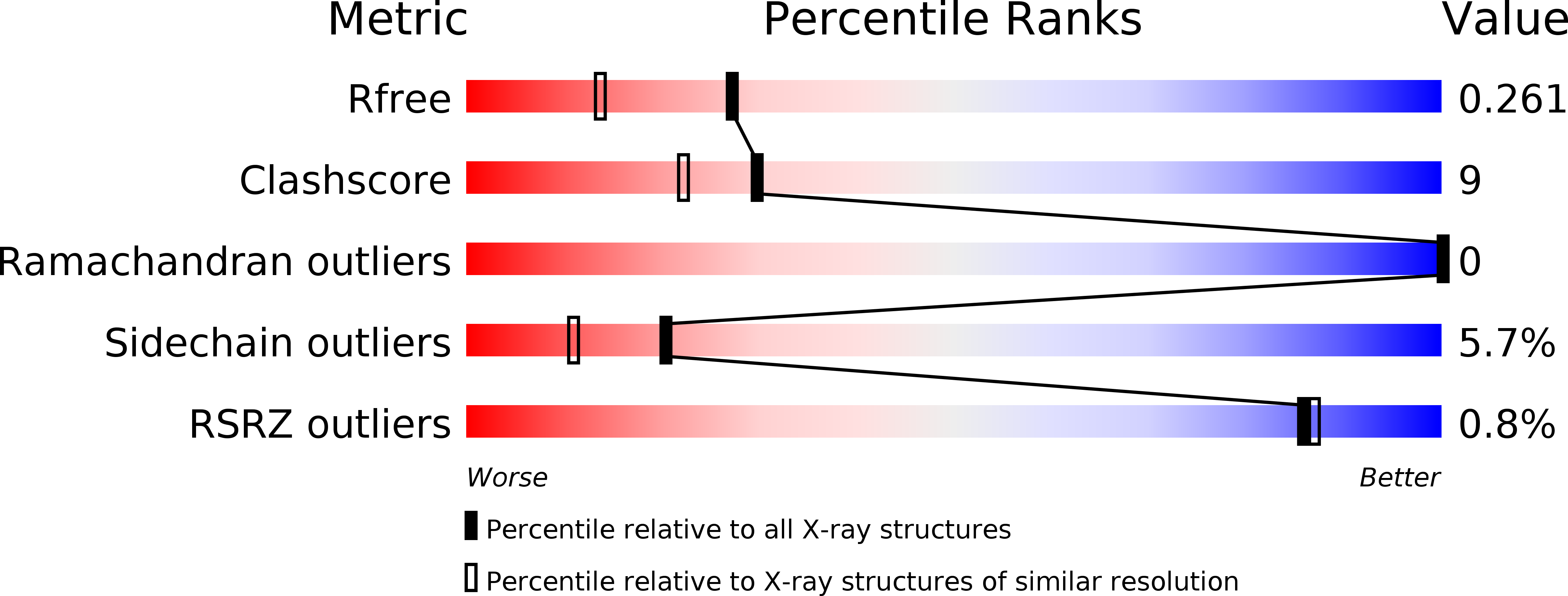
R-Value Free:
0.26
R-Value Work:
0.19
R-Value Observed:
0.20
Space Group:
P 65