3FYQ image
Deposition Date 2009-01-22
Release Date 2009-02-03
Last Version Date 2024-11-20
Entry Detail
PDB ID:
3FYQ
Keywords:
Title:
Structure of Drosophila melanogaster talin IBS2 domain (residues 1981-2168)
Biological Source:
Source Organism:
Host Organism:
Method Details:
Experimental Method:
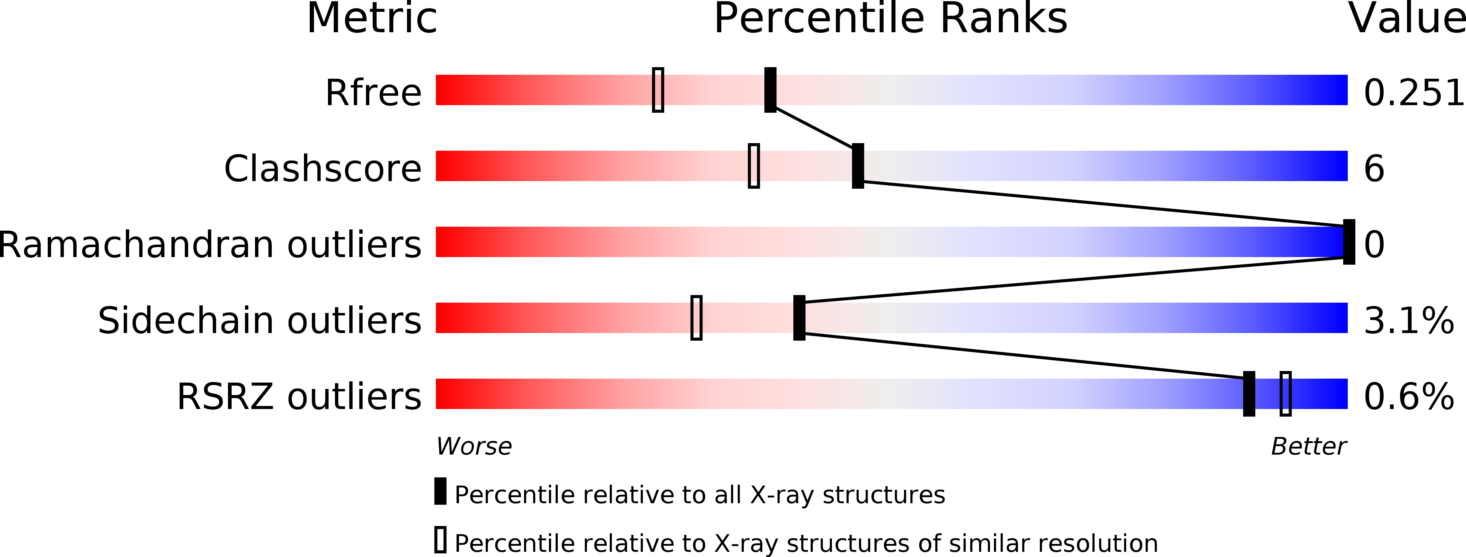
Resolution:
1.95 Å
R-Value Free:
0.25
R-Value Work:
0.20
R-Value Observed:
0.20
Space Group:
C 1 2 1
Macromolecular Entities
Structures with similar UniProt ID
Protein Blast
Polymer Type:polypeptide(L)
Molecule:CG6831-PA (Talin)
Gene (Uniprot):rhea
Chain IDs:A
Chain Length:199
Number of Molecules:1
Biological Source:Drosophila melanogaster
Modified Residue
Compound ID Chain ID Parent Comp ID Details 2D Image
MSE A MET SELENOMETHIONINE
Primary Citation
Crystal structure of the talin integrin binding domain 2.
J.Mol.Biol. 387 787 793 (2009)
PMID: 19340939

Abstact

Integrins are transmembrane receptors that mediate cell adhesion to the extracellular matrix and play essential roles in tissue development and maintenance. The cytoplasmic segment of integrin associates with talin, a large intracellular protein that links integrin to the actin cytoskeleton. Binding of talin via an integrin binding segment (IBS1) results in large conformational changes in the extracellular portion of integrin, which modulates the affinity of integrins for their extracellular matrix ligands. However, integrin binding also requires a second segment of talin (IBS2). Despite detailed descriptions of the integrin-IBS1 binding, the molecular determinants that drive the integrin-IBS2 association are poorly understood. Here, we describe the crystal structure of the talin IBS2 domain, which forms a five-helix bundle. The large structural homology with a vinculin binding domain hints at an ancient gene duplication and suggests that helix 4 may bind to vinculin if the bundle is unfolded. Mapping previous mutations on the surface highlights a likely binding interface for integrin.

Legend

Protein

Chemical

Disease

Primary Citation of related structures