3FXI image
Deposition Date 2009-01-21
Release Date 2009-03-03
Last Version Date 2024-11-06
Entry Detail
PDB ID:
3FXI
Keywords:
Title:
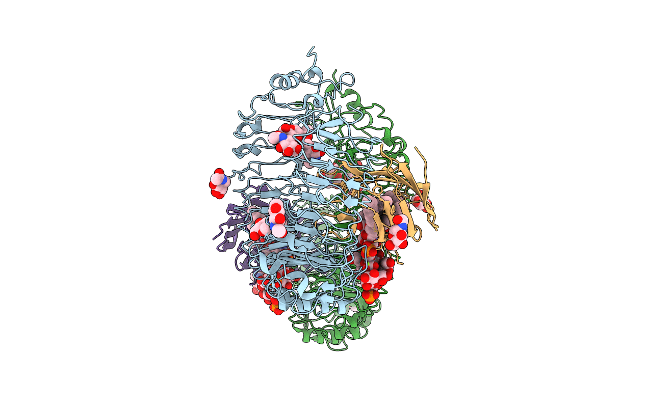
Crystal structure of the human TLR4-human MD-2-E.coli LPS Ra complex
Biological Source:
Source Organism:
Homo sapiens (Taxon ID: 9606)
Host Organism:
Method Details:
Experimental Method:
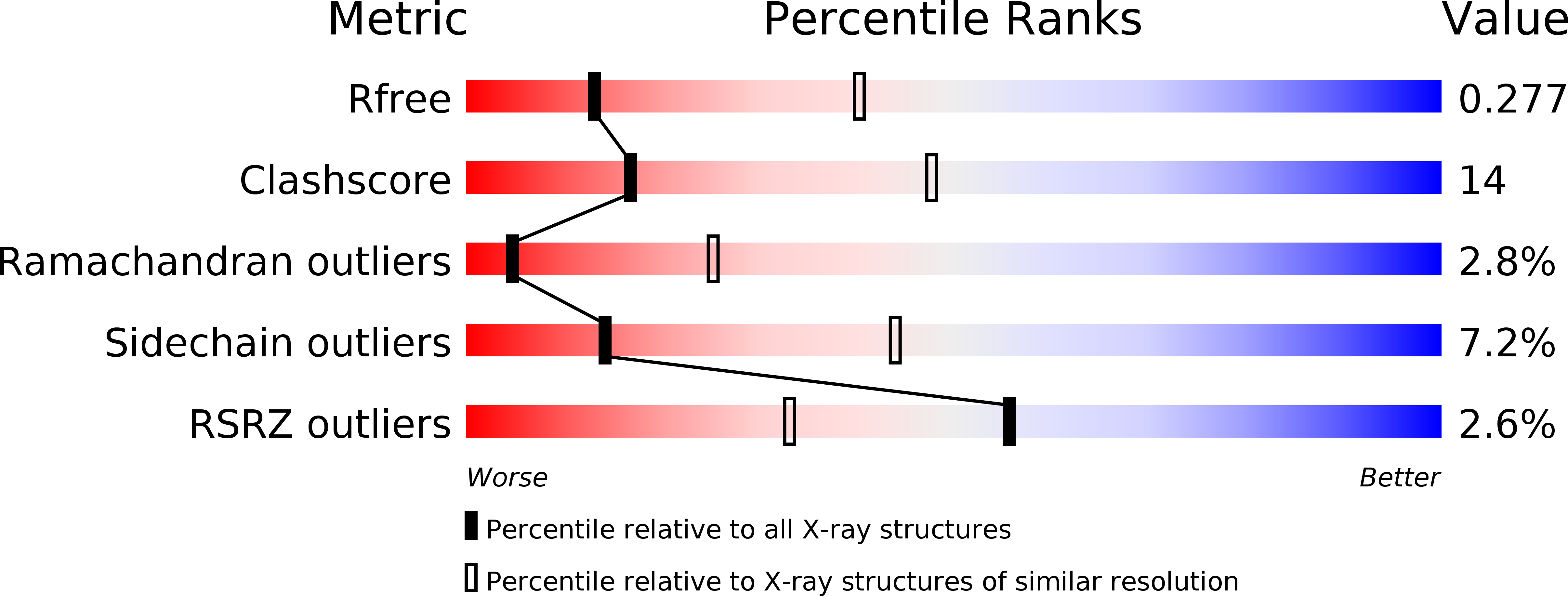
Resolution:
3.10 Å
R-Value Free:
0.28
R-Value Work:
0.24
Space Group:
P 21 21 21
Macromolecular Entities
Polymer Type:polypeptide(L)
Molecule:Toll-like receptor 4
Gene (Uniprot):TLR4
Chain IDs:A, B
Chain Length:605
Number of Molecules:2
Biological Source:Homo sapiens
Polymer Type:polypeptide(L)
Molecule:Lymphocyte antigen 96
Gene (Uniprot):LY96
Chain IDs:C, D
Chain Length:142
Number of Molecules:2
Biological Source:Homo sapiens
Modified Residue
Compound ID Chain ID Parent Comp ID Details 2D Image
ASN A ASN GLYCOSYLATION SITE
Primary Citation
The structural basis of lipopolysaccharide recognition by the TLR4-MD-2 complex
Nature 458 1191 1195 (2009)
PMID: 19252480 DOI: 10.1038/nature07830

Abstact

The lipopolysaccharide (LPS) of Gram negative bacteria is a well-known inducer of the innate immune response. Toll-like receptor (TLR) 4 and myeloid differentiation factor 2 (MD-2) form a heterodimer that recognizes a common 'pattern' in structurally diverse LPS molecules. To understand the ligand specificity and receptor activation mechanism of the TLR4-MD-2-LPS complex we determined its crystal structure. LPS binding induced the formation of an m-shaped receptor multimer composed of two copies of the TLR4-MD-2-LPS complex arranged symmetrically. LPS interacts with a large hydrophobic pocket in MD-2 and directly bridges the two components of the multimer. Five of the six lipid chains of LPS are buried deep inside the pocket and the remaining chain is exposed to the surface of MD-2, forming a hydrophobic interaction with the conserved phenylalanines of TLR4. The F126 loop of MD-2 undergoes localized structural change and supports this core hydrophobic interface by making hydrophilic interactions with TLR4. Comparison with the structures of tetra-acylated antagonists bound to MD-2 indicates that two other lipid chains in LPS displace the phosphorylated glucosamine backbone by approximately 5 A towards the solvent area. This structural shift allows phosphate groups of LPS to contribute to receptor multimerization by forming ionic interactions with a cluster of positively charged residues in TLR4 and MD-2. The TLR4-MD-2-LPS structure illustrates the remarkable versatility of the ligand recognition mechanisms employed by the TLR family, which is essential for defence against diverse microbial infection.

Legend

Protein

Chemical

Disease

Primary Citation of related structures