3FWL image
Deposition Date 2009-01-19
Release Date 2009-06-02
Last Version Date 2024-10-30
Entry Detail
PDB ID:
3FWL
Title:
Crystal Structure of the Full-Length Transglycosylase PBP1b from Escherichia coli
Biological Source:
Source Organism:
Escherichia coli (Taxon ID: 562)
Host Organism:
Method Details:
Experimental Method:
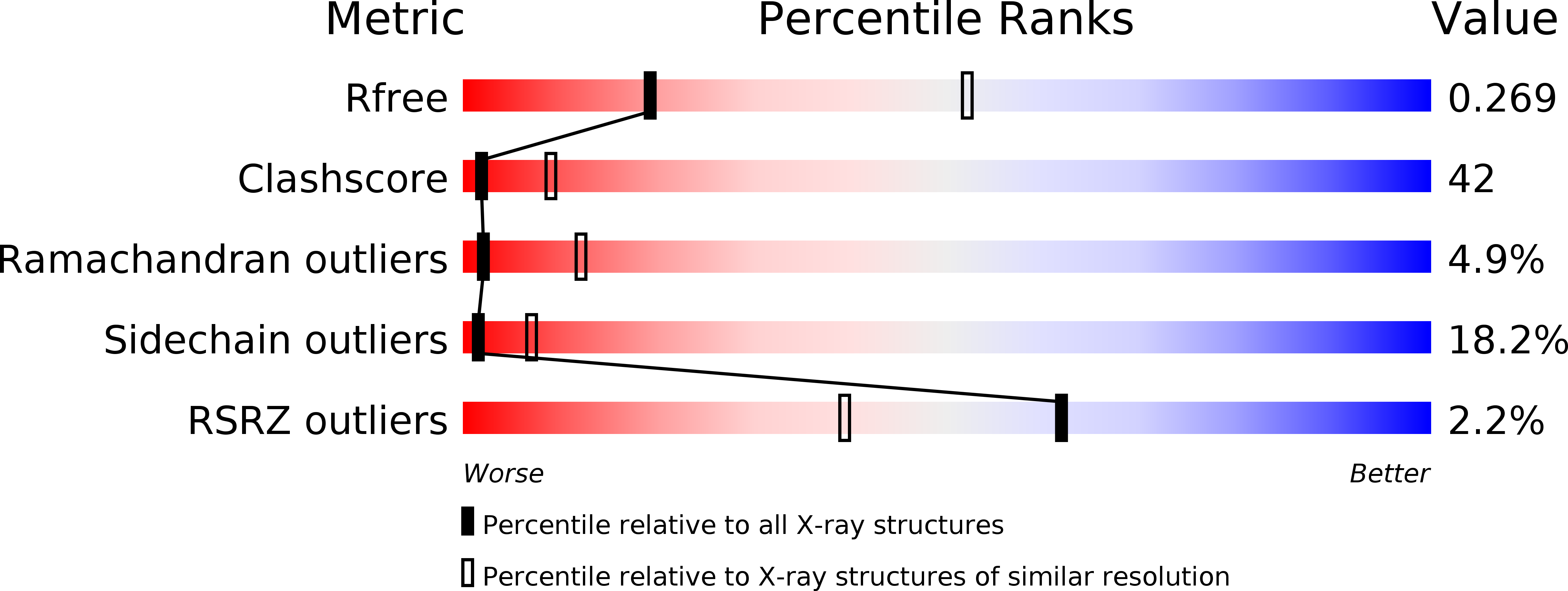
Resolution:
3.09 Å
R-Value Free:
0.27
R-Value Work:
0.21
R-Value Observed:
0.21
Space Group:
P 21 21 2
Macromolecular Entities
Polymer Type:polypeptide(L)
Molecule:Penicillin-binding protein 1B
Gene (Uniprot):mrcB
Chain IDs:A
Chain Length:751
Number of Molecules:1
Biological Source:Escherichia coli
Modified Residue
Compound ID Chain ID Parent Comp ID Details 2D Image
MSE A MET SELENOMETHIONINE
Ligand Molecules
Primary Citation
Crystal structure of the membrane-bound bifunctional transglycosylase PBP1b from Escherichia coli.
Proc.Natl.Acad.Sci.USA 106 8824 8829 (2009)
PMID: 19458048 DOI: 10.1073/pnas.0904030106

Abstact

Drug-resistant bacteria have caused serious medical problems in recent years, and the need for new antibacterial agents is undisputed. Transglycosylase, a multidomain membrane protein essential for cell wall synthesis, is an excellent target for the development of new antibiotics. Here, we determined the X-ray crystal structure of the bifunctional transglycosylase penicillin-binding protein 1b (PBP1b) from Escherichia coli in complex with its inhibitor moenomycin to 2.16-A resolution. In addition to the transglycosylase and transpeptidase domains, our structure provides a complete visualization of this important antibacterial target, and reveals a domain for protein-protein interaction and a transmembrane helix domain essential for substrate binding, enzymatic activity, and membrane orientation.

Legend

Protein

Chemical

Disease

Primary Citation of related structures