3FUS image
Entry Detail
PDB ID:
3FUS
Keywords:
Title:
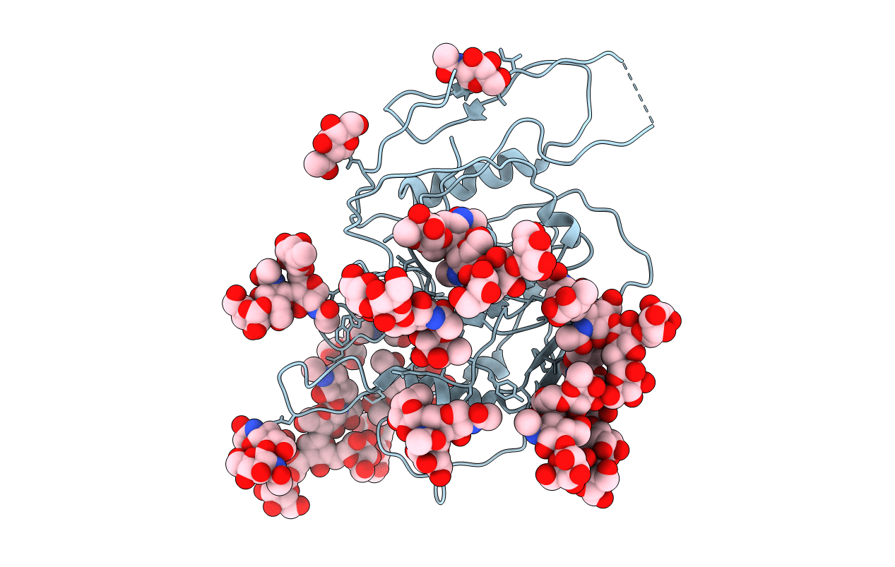
Improved Structure of the Unliganded Simian Immunodeficiency Virus gp120 Core
Biological Source:
Host Organism:
PDB Version:
Deposition Date:
2009-01-14
Release Date:
2009-06-30
Method Details:
Experimental Method:
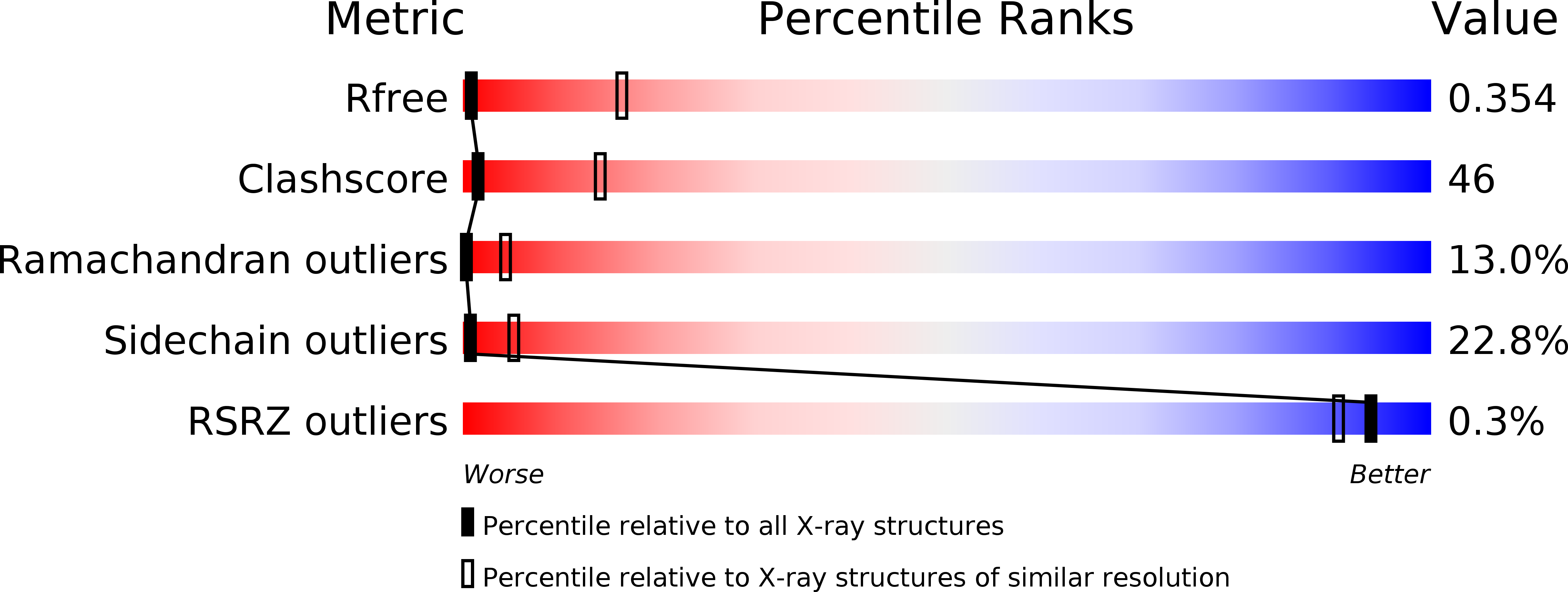
Resolution:
4.00 Å
R-Value Free:
0.35
R-Value Work:
0.34
R-Value Observed:
0.34
Space Group:
P 43 21 2
Macromolecular Entities
Polymer Type:polypeptide(L)
Description:EXTERIOR MEMBRANE GLYCOPROTEIN GP120
Chain IDs:A
Chain Length:316
Number of Molecules:1
Biological Source:Simian immunodeficiency virus
Modified Residue
Compound ID Chain ID Parent Comp ID Details 2D Image
ASN A ASN GLYCOSYLATION SITE
Ligand Molecules
Primary Citation
Structural improvement of unliganded simian immunodeficiency virus gp120 core by normal-mode-based X-ray crystallographic refinement.
Acta Crystallogr.,Sect.D 65 339 347 (2009)
PMID: 19307715 DOI: 10.1107/S0907444909003539

Abstact

The envelope protein gp120/gp41 of simian and human immunodeficiency viruses plays a critical role in viral entry into host cells. However, the extraordinarily high structural flexibility and heavy glycosylation of the protein have presented enormous difficulties in the pursuit of high-resolution structural investigation of some of its conformational states. An unliganded and fully glycosylated gp120 core structure was recently determined to 4.0 A resolution. The rather low data-to-parameter ratio limited refinement efforts in the original structure determination. In this work, refinement of this gp120 core structure was carried out using a normal-mode-based refinement method that has been shown in previous studies to be effective in improving models of a supramolecular complex at 3.42 A resolution and of a membrane protein at 3.2 A resolution. By using only the first four nonzero lowest-frequency normal modes to construct the anisotropic thermal parameters, combined with manual adjustments and standard positional refinement using REFMAC5, the structural model of the gp120 core was significantly improved in many aspects, including substantial decreases in R factors, better fitting of several flexible regions in electron-density maps, the addition of five new sugar rings at four glycan chains and an excellent correlation of the B-factor distribution with known structural flexibility. These results further underscore the effectiveness of this normal-mode-based method in improving models of protein and nonprotein components in low-resolution X-ray structures.

Legend

Protein

Chemical

Disease

Primary Citation of related structures