Deposition Date
2009-01-14
Release Date
2009-06-02
Last Version Date
2024-10-30
Entry Detail
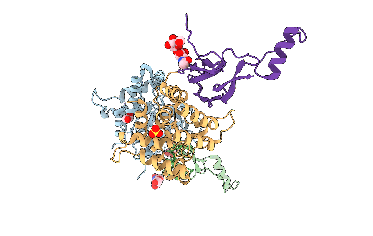
Biological Source:
Source Organism(s):
Homo sapiens (Taxon ID: 9606)
Rattus norvegicus (Taxon ID: 10116)
Rattus norvegicus (Taxon ID: 10116)
Expression System(s):
Method Details:
Experimental Method:
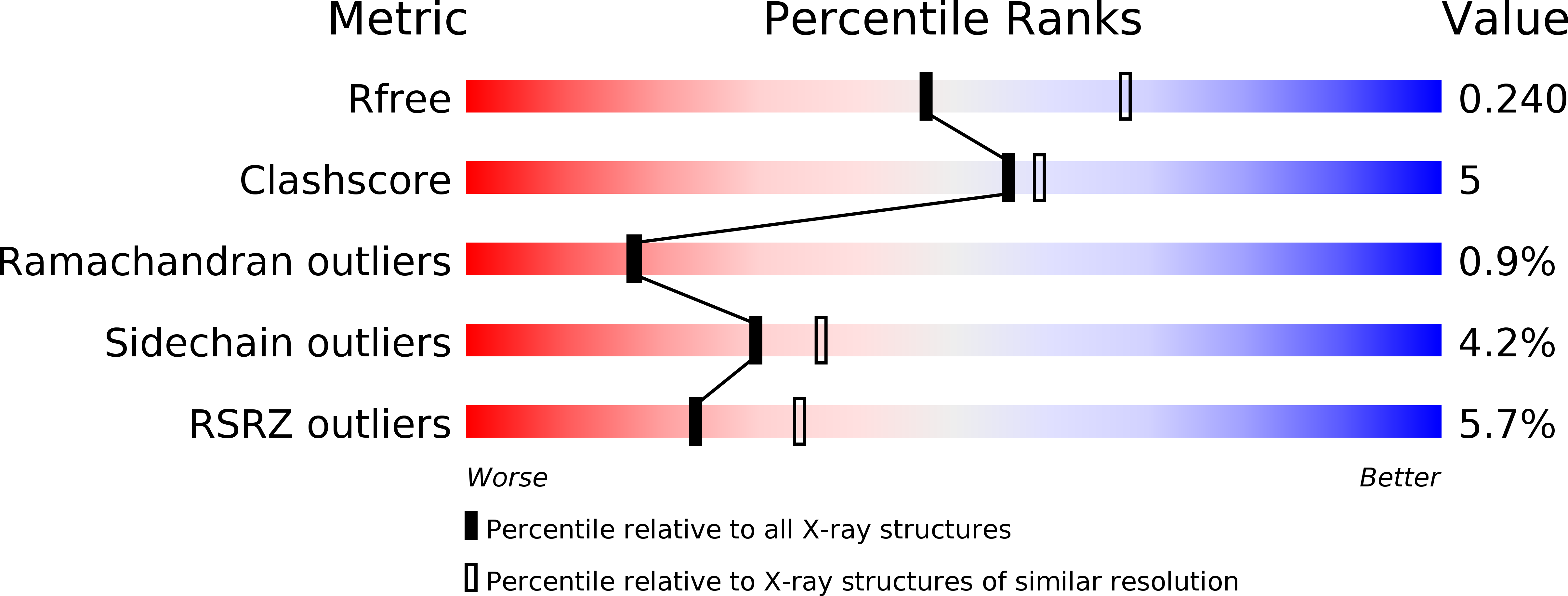
Resolution:
2.35 Å
R-Value Free:
0.28
R-Value Work:
0.22
R-Value Observed:
0.22
Space Group:
C 1 2 1