3FTC image
Deposition Date 2009-01-12
Release Date 2009-03-24
Last Version Date 2024-11-20
Entry Detail
PDB ID:
3FTC
Keywords:
Title:
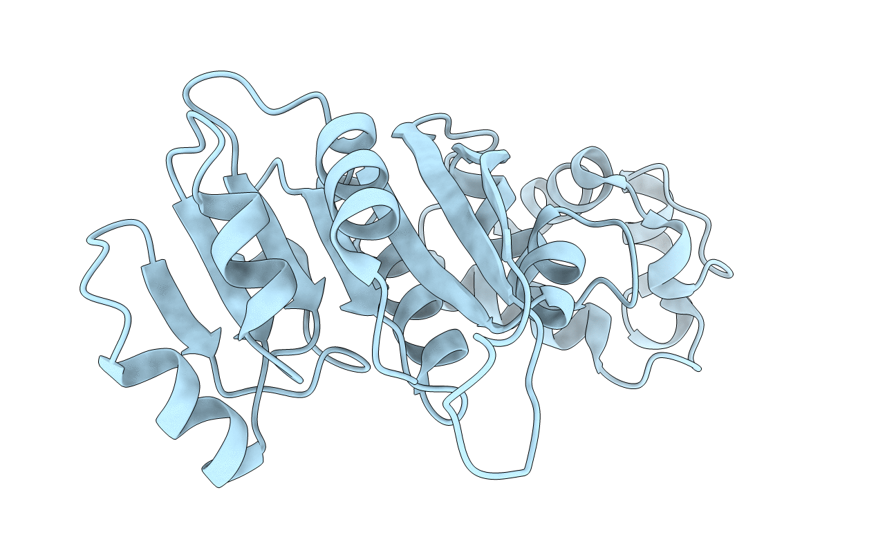
Crystal structure of A. aeolicus KsgA at 1.72-Angstrom resolution
Biological Source:
Source Organism:
Host Organism:
Method Details:
Experimental Method:
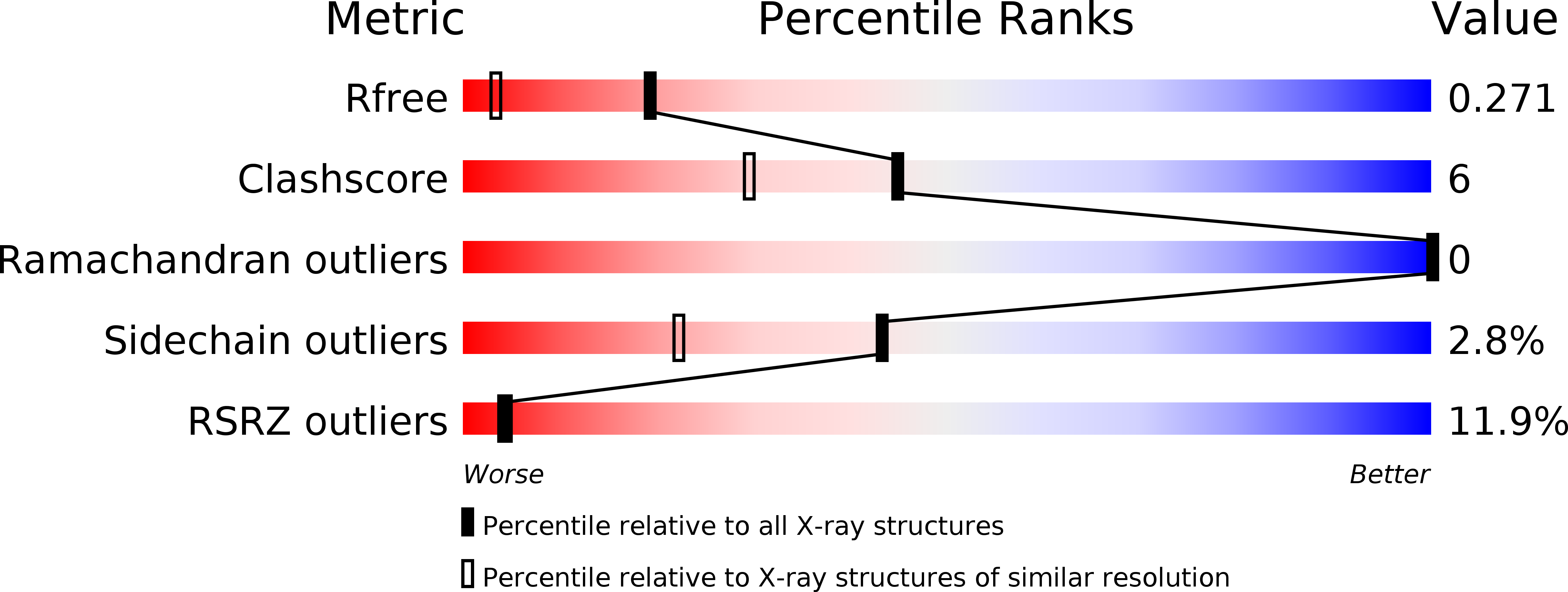
Resolution:
1.68 Å
R-Value Free:
0.24
R-Value Work:
0.20
R-Value Observed:
0.20
Space Group:
P 21 21 21
Macromolecular Entities
Polymer Type:polypeptide(L)
Molecule:Dimethyladenosine transferase
Gene (Uniprot):rsmA
Chain IDs:A
Chain Length:249
Number of Molecules:1
Biological Source:Aquifex aeolicus
Primary Citation
Structural Basis for Binding of RNA and Cofactor by a KsgA Methyltransferase.
Structure 17 374 385 (2009)
PMID: 19278652 DOI: 10.1016/j.str.2009.01.010

Abstact

Among methyltransferases, KsgA and the reaction it catalyzes are conserved throughout evolution. However, the specifics of substrate recognition by the enzyme remain unknown. Here we report structures of Aquifex aeolicus KsgA, in its ligand-free form, in complex with RNA, and in complex with both RNA and S-adenosylhomocysteine (SAH, reaction product of cofactor S-adenosylmethionine), revealing critical structural information on KsgA-RNA and KsgA-SAH interactions. Moreover, the structures show how conformational changes that occur upon RNA binding create the cofactor-binding site. There are nine conserved functional motifs (motifs I-VIII and X) in KsgA. Prior to RNA binding, motifs I and VIII are flexible, each exhibiting two distinct conformations. Upon RNA binding, the two motifs become stabilized in one of these conformations, which is compatible with the binding of SAH. Motif X, which is also stabilized upon RNA binding, is directly involved in the binding of SAH.

Legend

Protein

Chemical

Disease

Primary Citation of related structures