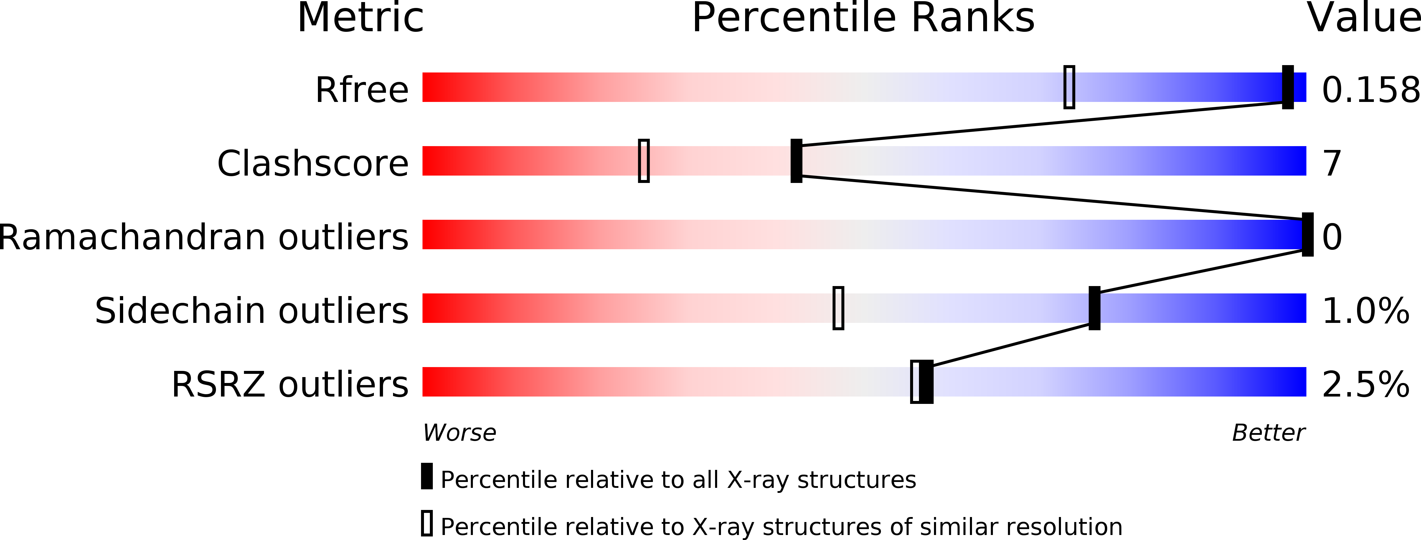
3FRH image
Deposition Date 2009-01-08
Release Date 2009-08-11
Last Version Date 2023-11-01
Entry Detail
PDB ID:
3FRH
Keywords:
Title:
Structure of the 16S rRNA methylase RmtB, P21
Biological Source:
Source Organism:
Escherichia coli (Taxon ID: 562)
Host Organism:
Method Details:
Experimental Method:
Resolution:
1.20 Å
R-Value Free:
0.15
R-Value Work:
0.13
R-Value Observed:
0.13
Space Group:
P 1 21 1
Macromolecular Entities
Structures with similar UniProt ID
Protein Blast
Polymer Type:polypeptide(L)
Molecule:16S rRNA methylase
Gene (Uniprot):rmtB
Chain IDs:A
Chain Length:253
Number of Molecules:1
Biological Source:Escherichia coli
Ligand Molecules
Primary Citation
Structural bases for 16 S rRNA methylation catalyzed by ArmA and RmtB methyltransferases
J.Mol.Biol. 388 570 582 (2009)
PMID: 19303884 DOI: 10.1016/j.jmb.2009.03.034

Abstact

Aminoglycosides are used extensively for the treatment of severe infections due to Gram-negative bacteria. However, certain species have become highly resistant after acquisition of genes for methyltransferases which catalyze post-transcriptional methylation of N7-G1405 in 16 S rRNA of 30 S ribosomal subunits. Inactivation of this enzymatic activity is therefore an important challenge for development of an effective therapy. The present work describes the crystallographic structures of methyltransferases RmtB and ArmA from clinical isolates. Together with biochemical experiments, the 3D structures indicate that the N-terminal domain specific for this family of methyltransferases is required for enzymatic activity. Site-directed mutagenesis has enabled important residues for catalysis and RNA binding to be identified. These high-resolution structures should underpin the design of potential inhibitors of these enzymes, which could be used to restore the activity of aminoglycosides against resistant pathogens.

Legend

Protein

Chemical

Disease

Primary Citation of related structures