3FR7 image
Deposition Date 2009-01-08
Release Date 2009-04-14
Last Version Date 2023-09-06
Entry Detail
PDB ID:
3FR7
Keywords:
Title:
ketol-acid reductoisomerase (KARI) in complex with Mg2+
Biological Source:
Source Organism:
Host Organism:
Method Details:
Experimental Method:
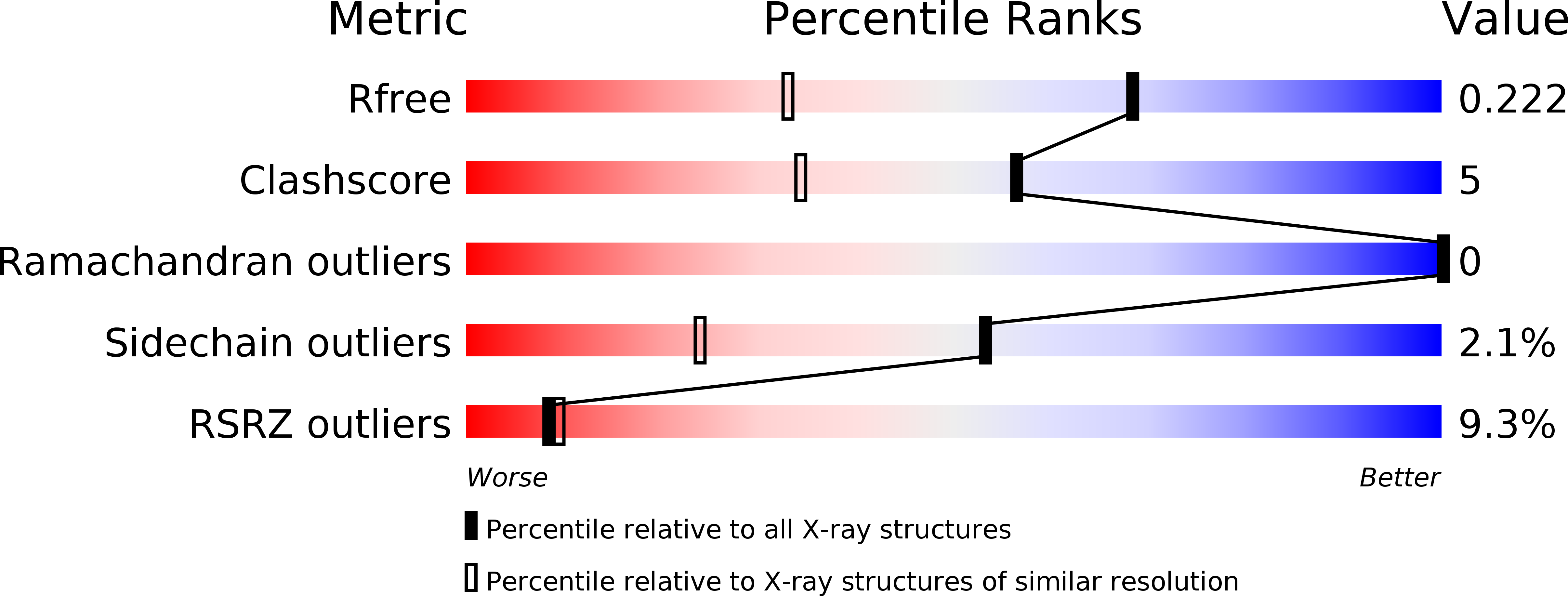
Resolution:
1.55 Å
R-Value Free:
0.23
R-Value Work:
0.20
R-Value Observed:
0.20
Space Group:
C 1 2 1
Macromolecular Entities
Structures with similar UniProt ID
Protein Blast
Polymer Type:polypeptide(L)
Molecule:Putative ketol-acid reductoisomerase (Os05g0573700 protein)
Gene (Uniprot):OJ1735_C10.18
Chain IDs:A, B
Chain Length:525
Number of Molecules:2
Biological Source:Oryza sativa Japonica Group
Ligand Molecules
Primary Citation
Conformational changes in a plant ketol-acid reductoisomerase upon Mg(2+) and NADPH binding as revealed by two crystal structures
J.Mol.Biol. 389 167 182 (2009)
PMID: 19362563 DOI: 10.1016/j.jmb.2009.04.012

Abstact

Ketol-acid reductoisomerase (KARI; EC 1.1.1.86) is an enzyme in the branched-chain amino acid biosynthesis pathway where it catalyzes the conversion of 2-acetolactate into (2R)-2,3-dihydroxy-3-isovalerate or the conversion of 2-aceto-2-hydroxybutyrate into (2R,3R)-2,3-dihydroxy-3-methylvalerate. KARI catalyzes two reactions-alkyl migration and reduction-and requires Mg(2+) and NADPH for activity. To date, the only reported structures for a plant KARI are those of the spinach enzyme-Mn(2+)-(phospho)ADP ribose-(2R,3R)-2,3-dihydroxy-3-methylvalerate complex and the spinach KARI-Mg(2)(+)-NADPH-N-hydroxy-N-isopropyloxamate complex, where N-hydroxy-N-isopropyloxamate is a predicted transition-state analog. These studies demonstrated that the enzyme consists of two domains, N-domain and C-domain, with the active site at the interface of these domains. Here, we have determined the structures of the rice KARI-Mg(2+) and rice KARI-Mg(2)(+)-NADPH complexes to 1.55 A and 2.80 A resolutions, respectively. In comparing the structures of all the complexes, several differences are observed. Firstly, the N-domain is rotated up to 15 degrees relative to the C-domain, expanding the active site by up to 4 A. Secondly, an alpha-helix in the C-domain that includes residues V510-T519 and forms part of the active site moves by approximately 3.9 A upon binding of NADPH. Thirdly, the 15 C-terminal amino acid residues in the rice KARI-Mg(2+) complex are disordered. In the rice KARI-Mg(2)(+)-NADPH complex and the spinach KARI structures, many of the 15 residues bind to NADPH and the N-domain and cover the active site. Fourthly, the location of the metal ions within the active site can vary by up to 2.7 A. The new structures allow us to propose that an induced-fit mechanism operates to (i) allow substrate to enter the active site, (ii) close over the active site during catalysis, and (iii) open the active site to facilitate product release.

Legend

Protein

Chemical

Disease

Primary Citation of related structures
Feedback Form
Name
Email
Institute
Feedback