
Deposition Date
2009-01-07
Release Date
2009-03-10
Last Version Date
2023-09-06
Entry Detail
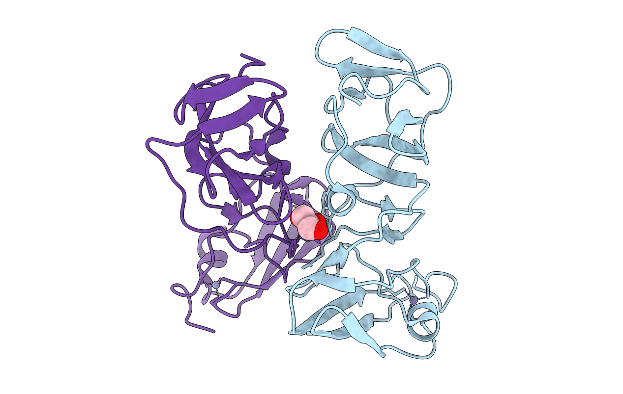
PDB ID:
3FQM
Keywords:
Title:
Crystal structure of a novel dimeric form of HCV NS5A domain I protein
Biological Source:
Source Organism(s):
Hepatitis C virus (isolate Con1) (Taxon ID: 333284)
Expression System(s):
Method Details:
Experimental Method:
Resolution:
1.90 Å
R-Value Free:
0.28
R-Value Work:
0.24
R-Value Observed:
0.24
Space Group:
P 31 2 1
