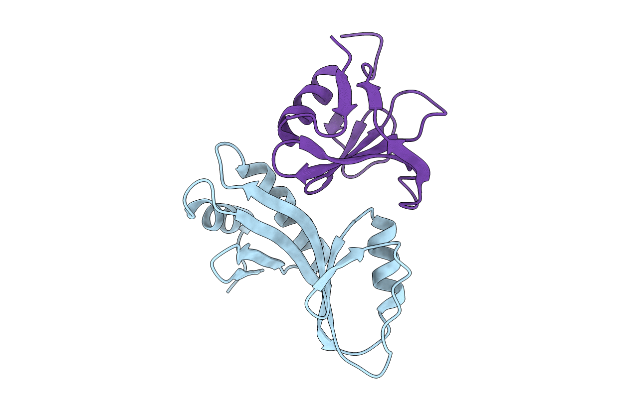
3FPN image
Deposition Date 2009-01-05
Release Date 2009-03-24
Last Version Date 2023-09-06
Entry Detail
PDB ID:
3FPN
Title:
Crystal structure of UvrA-UvrB interaction domains
Biological Source:
Source Organism:
Host Organism:
Method Details:
Experimental Method:
Resolution:
1.80 Å
R-Value Free:
0.24
R-Value Work:
0.23
R-Value Observed:
0.23
Space Group:
P 41 21 2
Macromolecular Entities
Polymer Type:polypeptide(L)
Molecule:Geobacillus stearothermophilus UvrA interaction domain
Chain IDs:A
Chain Length:119
Number of Molecules:1
Biological Source:Geobacillus stearothermophilus
Polymer Type:polypeptide(L)
Molecule:Geobacillus stearothermophilus UvrB interaction domain
Chain IDs:B
Chain Length:106
Number of Molecules:1
Biological Source:Geobacillus stearothermophilus
Primary Citation
A Structural Model for the Damage-sensing Complex in Bacterial Nucleotide Excision Repair.
J.Biol.Chem. 284 12837 12844 (2009)
PMID: 19287003 DOI: 10.1074/jbc.M900571200

Abstact

Nucleotide excision repair is distinguished from other DNA repair pathways by its ability to process a wide range of structurally unrelated DNA lesions. In bacteria, damage recognition is achieved by the UvrA.UvrB ensemble. Here, we report the structure of the complex between the interaction domains of UvrA and UvrB. These domains are necessary and sufficient for full-length UvrA and UvrB to associate and thereby form the DNA damage-sensing complex of bacterial nucleotide excision repair. The crystal structure and accompanying biochemical analyses suggest a model for the complete damage-sensing complex.

Legend

Protein

Chemical

Disease

Primary Citation of related structures