Deposition Date
2008-12-22
Release Date
2009-04-28
Last Version Date
2023-09-06
Entry Detail
Biological Source:
Source Organism(s):
Escherichia coli (Taxon ID: 83333)
Expression System(s):
Method Details:
Experimental Method:
Resolution:
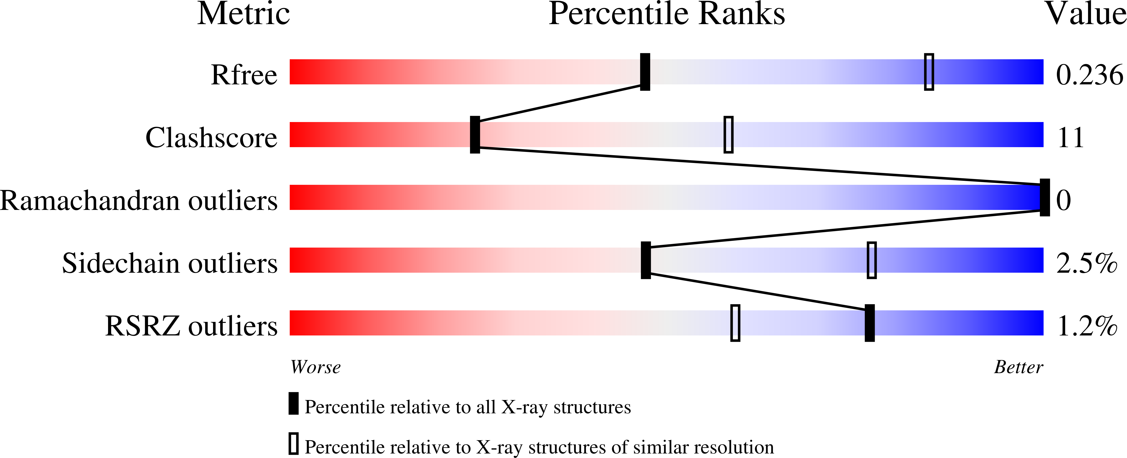
2.98 Å
R-Value Free:
0.24
R-Value Work:
0.21
R-Value Observed:
0.21
Space Group:
H 3