3FMF image
Entry Detail
PDB ID:
3FMF
Keywords:
Title:
Crystal structure of Mycobacterium tuberculosis dethiobiotin synthetase complexed with 7,8 diaminopelargonic acid carbamate
Biological Source:
Host Organism:
PDB Version:
Deposition Date:
2008-12-21
Release Date:
2009-06-30
Method Details:
Experimental Method:
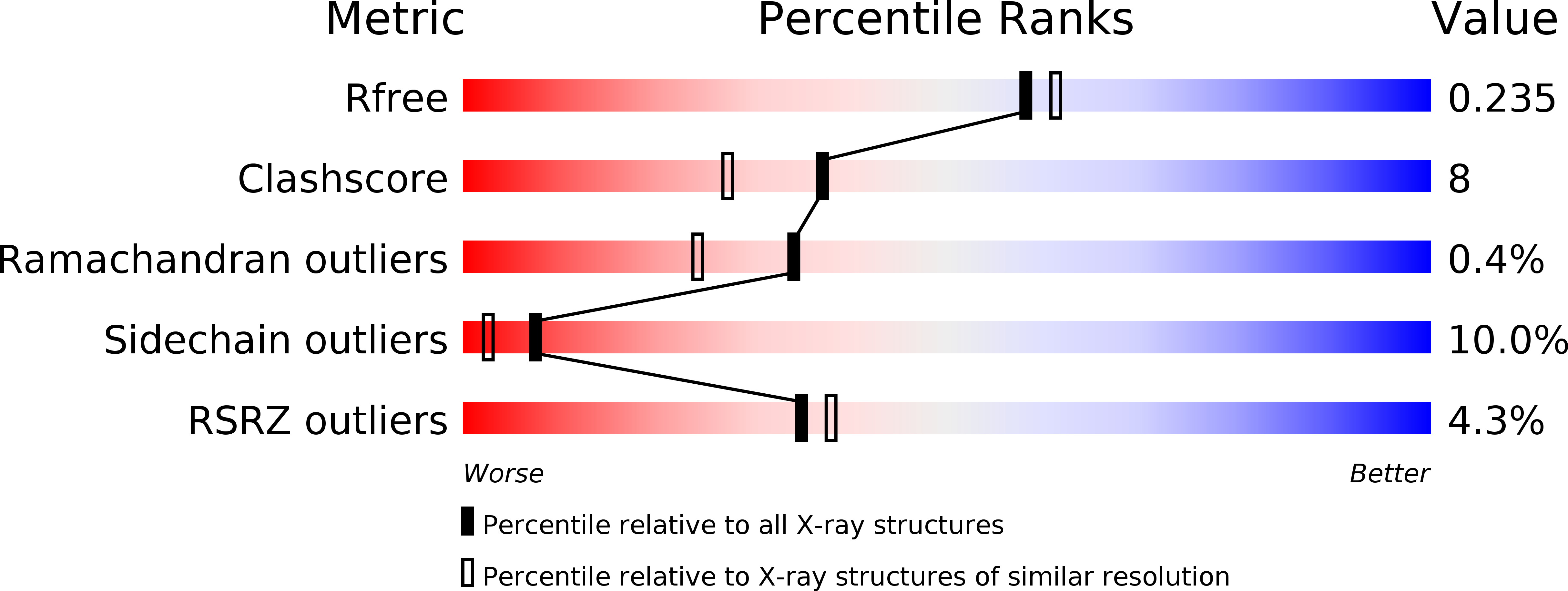
Resolution:
2.05 Å
R-Value Free:
0.23
R-Value Work:
0.19
R-Value Observed:
0.19
Space Group:
P 21 21 21
Macromolecular Entities
Polymer Type:polypeptide(L)
Description:Dethiobiotin synthetase
Chain IDs:A, B, C, D
Chain Length:251
Number of Molecules:4
Biological Source:Mycobacterium tuberculosis
Modified Residue
Compound ID Chain ID Parent Comp ID Details 2D Image
CSX A CYS S-OXY CYSTEINE
Ligand Molecules
Primary Citation
Structural characterization of the Mycobacterium tuberculosis biotin biosynthesis enzymes 7,8-diaminopelargonic acid synthase and dethiobiotin synthetase .
Biochemistry 49 6746 6760 (2010)
PMID: 20565114 DOI: 10.1021/bi902097j

Abstact

Mycobacterium tuberculosis (Mtb) depends on biotin synthesis for survival during infection. In the absence of biotin, disruption of the biotin biosynthesis pathway results in cell death rather than growth arrest, an unusual phenotype for an Mtb auxotroph. Humans lack the enzymes for biotin production, making the proteins of this essential Mtb pathway promising drug targets. To this end, we have determined the crystal structures of the second and third enzymes of the Mtb biotin biosynthetic pathway, 7,8-diaminopelargonic acid synthase (DAPAS) and dethiobiotin synthetase (DTBS), at respective resolutions of 2.2 and 1.85 A. Superimposition of the DAPAS structures bound either to the SAM analogue sinefungin or to 7-keto-8-aminopelargonic acid (KAPA) allowed us to map the putative binding site for the substrates and to propose a mechanism by which the enzyme accommodates their disparate structures. Comparison of the DTBS structures bound to the substrate 7,8-diaminopelargonic acid (DAPA) or to ADP and the product dethiobiotin (DTB) permitted derivation of an enzyme mechanism. There are significant differences between the Mtb enzymes and those of other organisms; the Bacillus subtilis DAPAS, presented here at a high resolution of 2.2 A, has active site variations and the Escherichia coli and Helicobacter pylori DTBS have alterations in their overall folds. We have begun to exploit the unique characteristics of the Mtb structures to design specific inhibitors against the biotin biosynthesis pathway in Mtb.

Legend

Protein

Chemical

Disease

Primary Citation of related structures