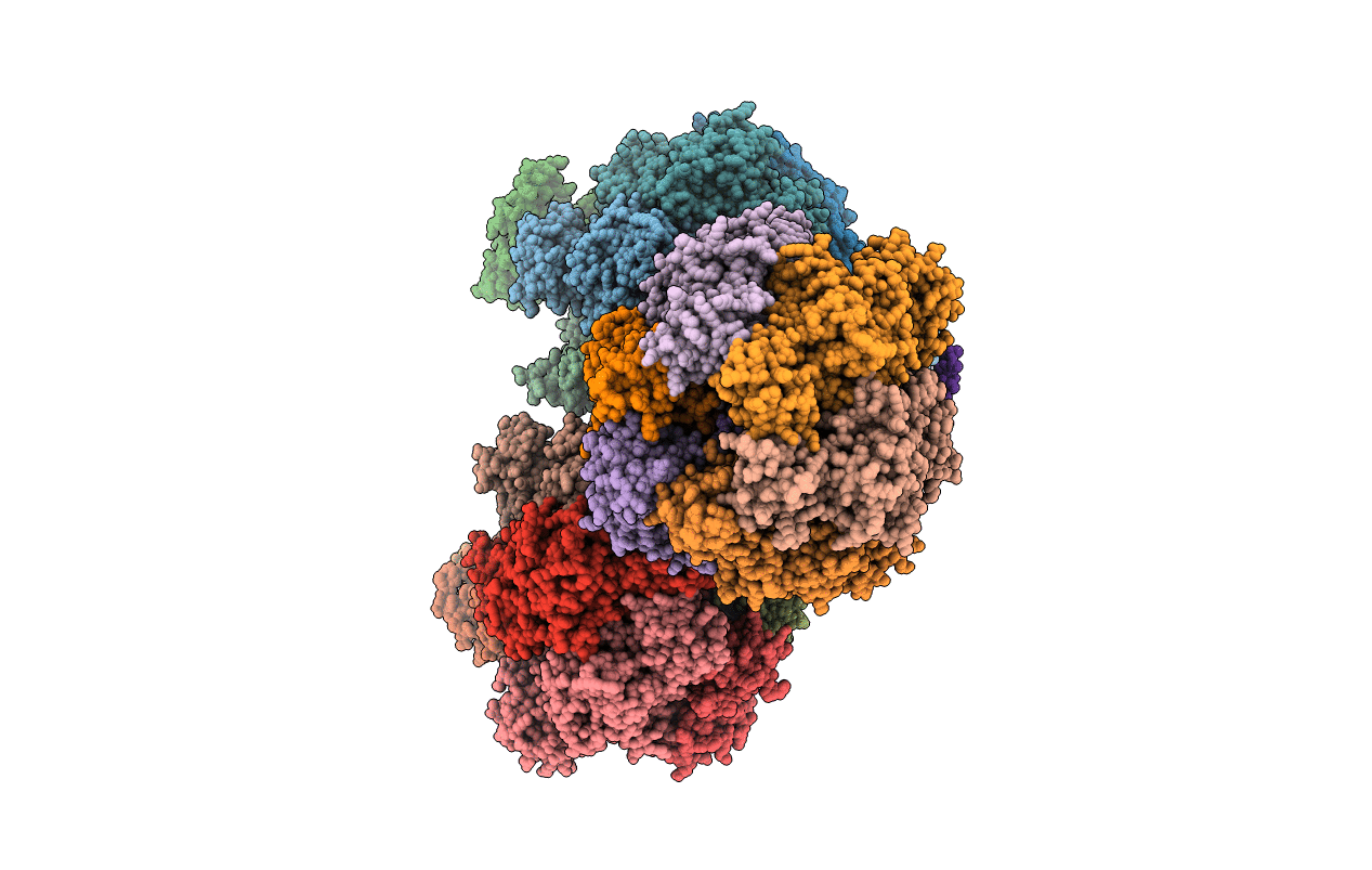
3FKS image
Deposition Date 2008-12-17
Release Date 2009-03-03
Last Version Date 2023-09-06
Entry Detail
PDB ID:
3FKS
Keywords:
Title:
Yeast F1 ATPase in the absence of bound nucleotides
Biological Source:
Source Organism:
Method Details:
Experimental Method:
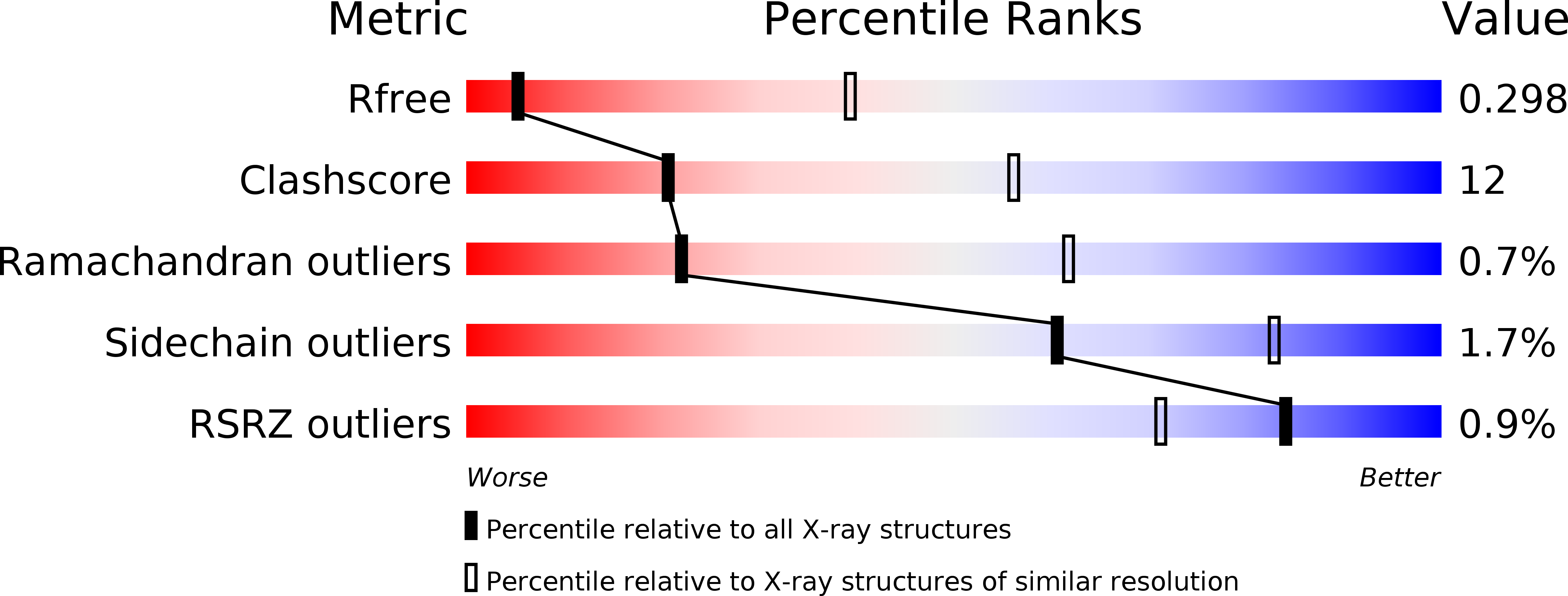
Resolution:
3.59 Å
R-Value Free:
0.30
R-Value Work:
0.24
R-Value Observed:
0.24
Space Group:
P 1 21 1
Macromolecular Entities
Polymer Type:polypeptide(L)
Molecule:ATP synthase subunit alpha, mitochondrial
Gene (Uniprot):ATP1
Chain IDs:A, B, C, J, K, L, S, T, U
Chain Length:510
Number of Molecules:9
Biological Source:Saccharomyces cerevisiae
Polymer Type:polypeptide(L)
Molecule:ATP synthase subunit beta, mitochondrial
Gene (Uniprot):ATP2
Chain IDs:D, E, F, M, N, O, V, W, X
Chain Length:484
Number of Molecules:9
Biological Source:Saccharomyces cerevisiae
Polymer Type:polypeptide(L)
Molecule:ATP synthase subunit gamma, mitochondrial
Gene (Uniprot):ATP3
Chain IDs:G, P, Y
Chain Length:278
Number of Molecules:3
Biological Source:Saccharomyces cerevisiae
Polymer Type:polypeptide(L)
Molecule:ATP synthase subunit delta, mitochondrial
Gene (Uniprot):ATP16
Chain IDs:H, Q, Z
Chain Length:138
Number of Molecules:3
Biological Source:Saccharomyces cerevisiae
Polymer Type:polypeptide(L)
Molecule:ATP synthase subunit epsilon, mitochondrial
Gene (Uniprot):ATP15
Chain IDs:I, R, AA (auth: 1)
Chain Length:61
Number of Molecules:3
Biological Source:Saccharomyces cerevisiae
Primary Citation
Asymmetric structure of the yeast f1 ATPase in the absence of bound nucleotides.
J.Biol.Chem. 284 10546 10551 (2009)
PMID: 19233840 DOI: 10.1074/jbc.M900544200

Abstact

The crystal structure of nucleotide-free yeast F(1) ATPase has been determined at a resolution of 3.6 A. The overall structure is very similar to that of the ground state enzyme. In particular, the beta(DP) and beta(TP) subunits both adopt the closed conformation found in the ground state structure despite the absence of bound nucleotides. This implies that interactions between the gamma and beta subunits are as important as nucleotide occupancy in determining the conformational state of the beta subunits. Furthermore, this result suggests that for the mitochondrial enzyme, there is no state of nucleotide occupancy that would result in more than one of the beta subunits adopting the open conformation. The adenine-binding pocket of the beta(TP) subunit is disrupted in the apoenzyme, suggesting that the beta(DP) subunit is responsible for unisite catalytic activity.

Legend

Protein

Chemical

Disease

Primary Citation of related structures