3FJQ image
Deposition Date 2008-12-15
Release Date 2009-08-04
Last Version Date 2024-11-20
Entry Detail
PDB ID:
3FJQ
Keywords:
Title:
Crystal structure of cAMP-dependent protein kinase catalytic subunit alpha in complex with peptide inhibitor PKI alpha (6-25)
Biological Source:
Source Organism:
Mus musculus (Taxon ID: 10090)
Host Organism:
Method Details:
Experimental Method:
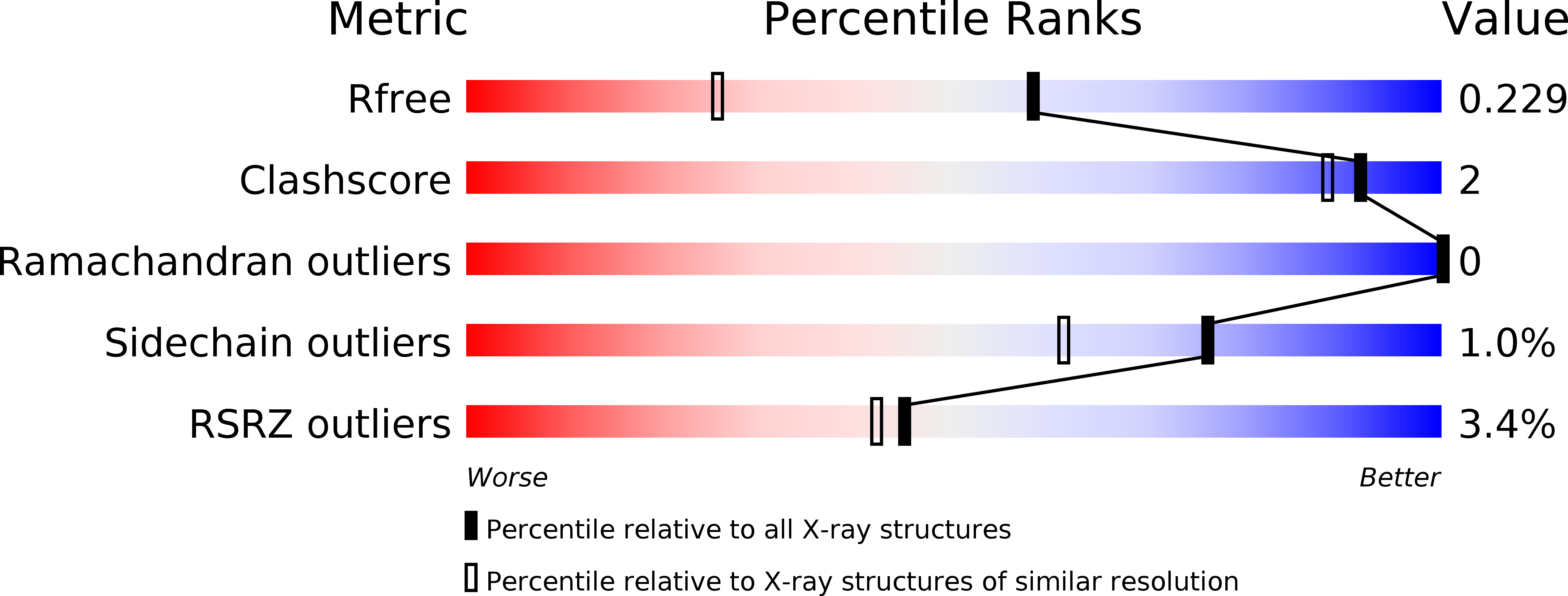
Resolution:
1.60 Å
R-Value Free:
0.20
R-Value Work:
0.17
R-Value Observed:
0.17
Space Group:
P 21 21 21
Macromolecular Entities
Structures with similar UniProt ID
Protein Blast
Polymer Type:polypeptide(L)
Molecule:cAMP-dependent protein kinase catalytic subunit alpha
Gene (Uniprot):Prkaca
Chain IDs:A (auth: E)
Chain Length:350
Number of Molecules:1
Biological Source:Mus musculus
Structures with similar UniProt ID
Protein Blast
Polymer Type:polypeptide(L)
Molecule:cAMP-dependent protein kinase inhibitor alpha
Gene (Uniprot):Pkia
Chain IDs:B (auth: I)
Chain Length:20
Number of Molecules:1
Biological Source:Mus musculus
Modified Residue
Compound ID Chain ID Parent Comp ID Details 2D Image
SEP A SER PHOSPHOSERINE
TPO A THR PHOSPHOTHREONINE
Primary Citation
Comparative surface geometry of the protein kinase family.
Protein Sci. 18 2016 2026 (2009)
PMID: 19610074 DOI: 10.1002/pro.209

Abstact

Identifying conserved pockets on the surfaces of a family of proteins can provide insight into conserved geometric features and sites of protein-protein interaction. Here we describe mapping and comparison of the surfaces of aligned crystallographic structures, using the protein kinase family as a model. Pockets are rapidly computed using two computer programs, FADE and Crevasse. FADE uses gradients of atomic density to locate grooves and pockets on the molecular surface. Crevasse, a new piece of software, splits the FADE output into distinct pockets. The computation was run on 10 kinase catalytic cores aligned on the alphaF-helix, and the resulting pockets spatially clustered. The active site cleft appears as a large, contiguous site that can be subdivided into nucleotide and substrate docking sites. Substrate specificity determinants in the active site cleft between serine/threonine and tyrosine kinases are visible and distinct. The active site clefts cluster tightly, showing a conserved spatial relationship between the active site and alphaF-helix in the C-lobe. When the alphaC-helix is examined, there are multiple mechanisms for anchoring the helix using spatially conserved docking sites. A novel site at the top of the N-lobe is present in all the kinases, and there is a large conserved pocket over the hinge and the alphaC-beta4 loop. Other pockets on the kinase core are strongly conserved but have not yet been mapped to a protein-protein interaction. Sites identified by this algorithm have revealed structural and spatially conserved features of the kinase family and potential conserved intermolecular and intramolecular binding sites.

Legend

Protein

Chemical

Disease

Primary Citation of related structures