3FJN image
Deposition Date 2008-12-14
Release Date 2009-11-24
Last Version Date 2024-11-06
Entry Detail
PDB ID:
3FJN
Keywords:
Title:
The crystal structure of 17-alpha hydroxysteroid dehydrogenase Y224D mutant.
Biological Source:
Source Organism:
Mus musculus (Taxon ID: 10090)
Host Organism:
Method Details:
Experimental Method:
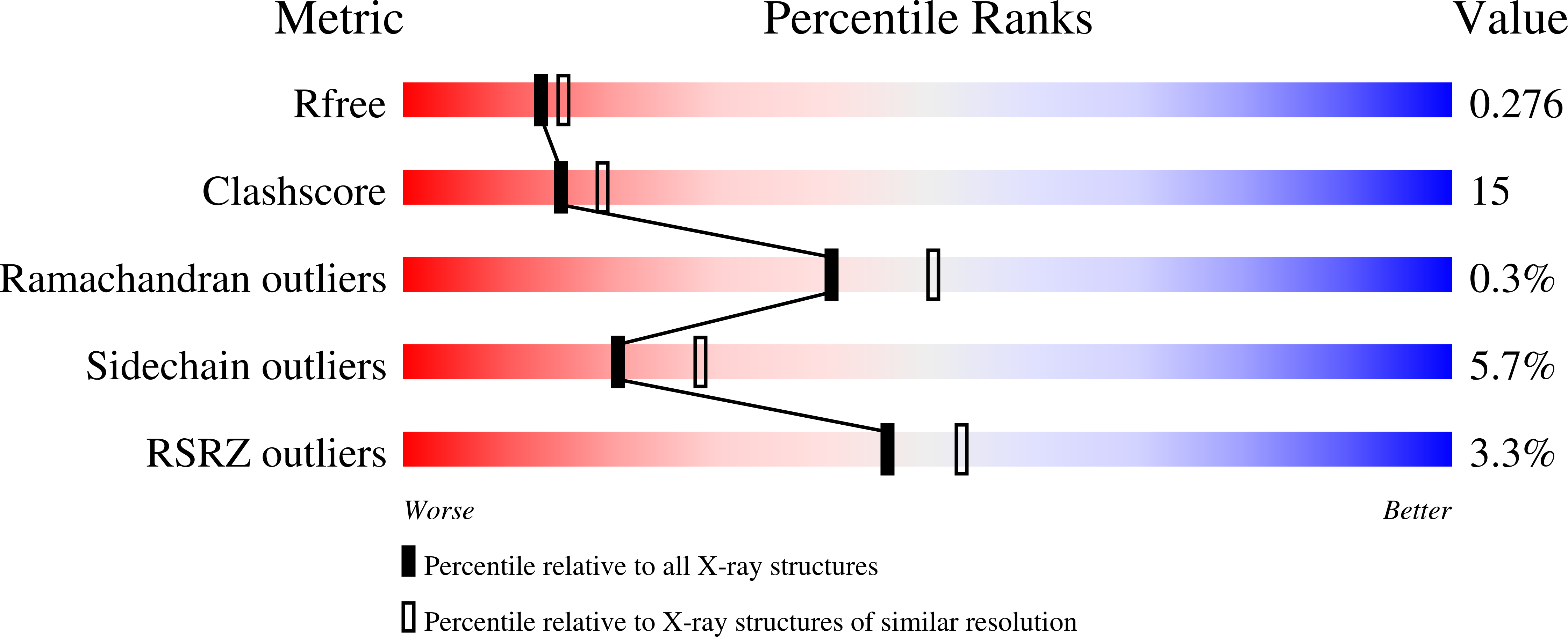
Resolution:
2.30 Å
R-Value Free:
0.27
R-Value Work:
0.17
R-Value Observed:
0.17
Space Group:
P 21 21 2
Macromolecular Entities
Structures with similar UniProt ID
Protein Blast
Polymer Type:polypeptide(L)
Molecule:Aldo-keto reductase family 1 member C21
Gene (Uniprot):Akr1c21
Mutagens:Y224D
Chain IDs:A, B
Chain Length:323
Number of Molecules:2
Biological Source:Mus musculus
Ligand Molecules
Primary Citation
Studies on a Tyr residue critical for the binding of coenzyme and substrate in mouse 3(17)alpha-hydroxysteroid dehydrogenase (AKR1C21): structure of the Y224D mutant enzyme
Acta Crystallogr.,Sect.D 66 198 204 (2010)
PMID: 20124700 DOI: 10.1107/S0907444909051464

Abstact

Mouse 3(17)alpha-hydroxysteroid dehydrogenase (AKR1C21) is the only aldo-keto reductase that catalyzes the stereospecific reduction of 3- and 17-ketosteroids to the corresponding 3(17)alpha-hydroxysteroids. The Y224D mutation of AKR1C21 reduced the K(m) value for NADP(H) by up to 80-fold and completely reversed the 17alpha stereospecificity of the enzyme. The crystal structure of the Y224D mutant at 2.3 A resolution revealed that the mutation resulted in a change in the conformation of the flexible loop B, including the V-shaped groove, which is a unique feature of the active-site architecture of wild-type AKR1C21 and is formed by the side chains of Tyr224 and Trp227. Furthermore, mutations (Y224F and Q222N) of residues involved in forming the safety belt for binding of the coenzyme showed similar alterations in kinetic constants for 3alpha-hydroxy/3-ketosteroids and 17-hydroxy/ketosteroids compared with the wild type.

Legend

Protein

Chemical

Disease

Primary Citation of related structures