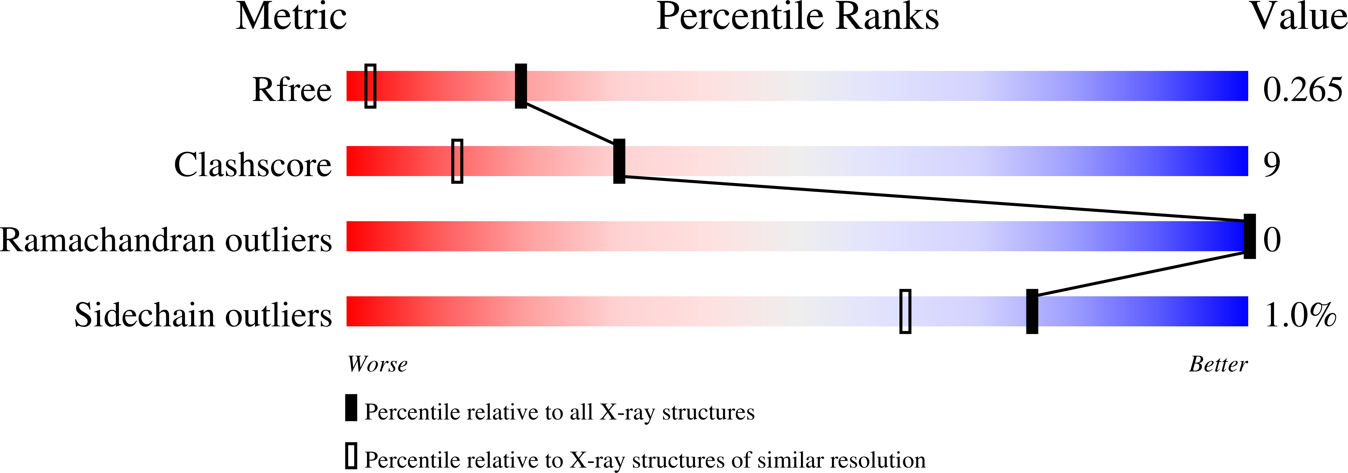
3FJ5 image
Deposition Date 2008-12-14
Release Date 2009-03-03
Last Version Date 2023-11-01
Entry Detail
PDB ID:
3FJ5
Keywords:
Title:
Crystal structure of the c-src-SH3 domain
Biological Source:
Source Organism:
Gallus gallus (Taxon ID: 9031)
Host Organism:
Method Details:
Experimental Method:
Resolution:
1.65 Å
R-Value Free:
0.22
R-Value Work:
0.20
R-Value Observed:
0.20
Space Group:
P 65
Macromolecular Entities
Polymer Type:polypeptide(L)
Molecule:Proto-oncogene tyrosine-protein kinase Src
Gene (Uniprot):SRC
Chain IDs:A, B
Chain Length:57
Number of Molecules:2
Biological Source:Gallus gallus
Primary Citation
Intertwined dimeric structure for the SH3 domain of the c-Src tyrosine kinase induced by polyethylene glycol binding
Febs Lett. 583 749 753 (2009)
PMID: 19185573 DOI: 10.1016/j.febslet.2009.01.036

Abstact

Here we report the first crystal structure of the SH3 domain of the cellular Src tyrosine kinase (c-Src-SH3) domain on its own. In the crystal two molecules of c-Src-SH3 exchange their -RT loops generating an intertwined dimer, in which the two SH3 units, preserving the binding site configuration, are oriented to allow simultaneous binding of two ligand molecules. The dimerization of c-Src-SH3 is induced, both in the crystal and in solution, by the binding of a PEG molecule at the dimer interface, indicating that this type of conformations are energetically close to the native structure. These results have important implications respect to in vivo oligomerization and amyloid aggregation.

Legend

Protein

Chemical

Disease

Primary Citation of related structures