3FHX image
Deposition Date 2008-12-10
Release Date 2008-12-23
Last Version Date 2023-09-06
Entry Detail
PDB ID:
3FHX
Keywords:
Title:
Crystal structure of D235A mutant of human pyridoxal kinase
Biological Source:
Source Organism:
Homo sapiens (Taxon ID: 9606)
Host Organism:
Method Details:
Experimental Method:
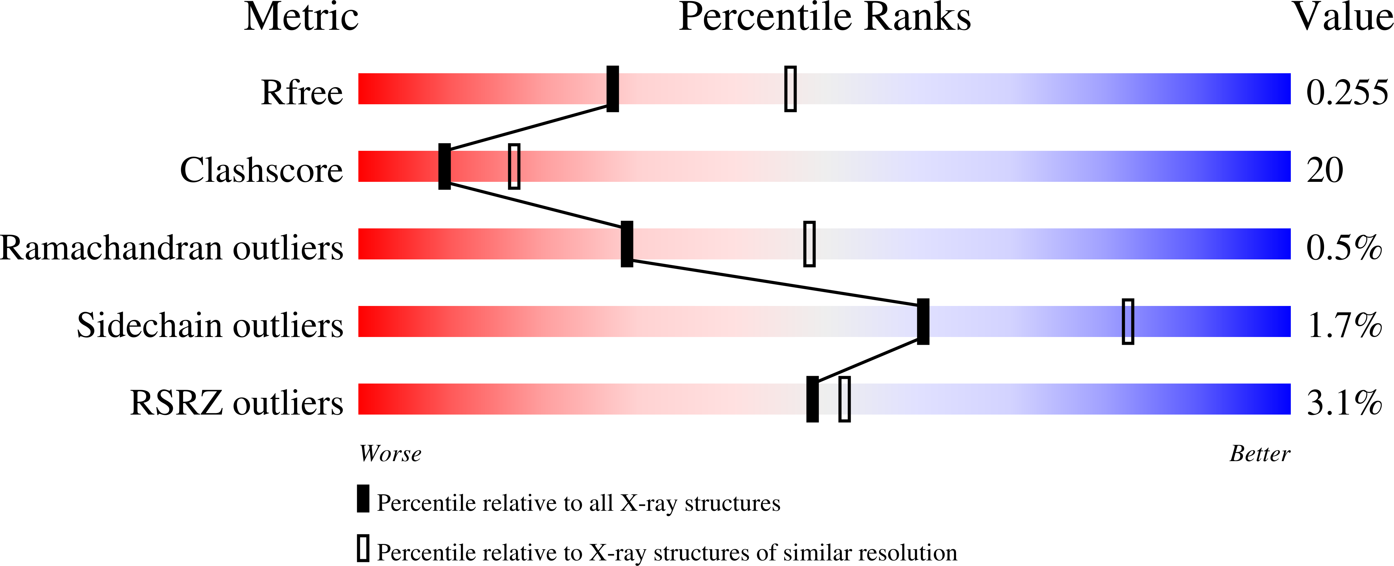
Resolution:
2.50 Å
R-Value Free:
0.26
R-Value Work:
0.21
R-Value Observed:
0.21
Space Group:
I 2 2 2
Macromolecular Entities
Polymer Type:polypeptide(L)
Molecule:Pyridoxal kinase
Gene (Uniprot):PDXK
Mutations:D235A
Chain IDs:A, B
Chain Length:312
Number of Molecules:2
Biological Source:Homo sapiens
Primary Citation
Kinetic and structural studies of the role of the active site residue Asp235 of human pyridoxal kinase.
Biochem.Biophys.Res.Commun. 381 12 15 (2009)
PMID: 19351586 DOI: 10.1016/j.bbrc.2009.01.170

Abstact

Pyridoxal kinase catalyzes the phosphorylation of pyridoxal (PL) to pyridoxal 5'-phosphate (PLP). A D235A variant shows 7-fold and 15-fold decreases in substrate affinity and activity, respectively. A D235N variant shows approximately 2-fold decrease in both PL affinity and activity. The crystal structure of D235A (2.5 A) shows bound ATP, PL and PLP, while D235N (2.3 A) shows bound ATP and sulfate. These results document the role of Asp235 in PL kinase activity. The observation that the active site of PL kinase can accommodate both ATP and PLP suggests that formation of a ternary Enz.PLP.ATP complex could occur in the wild-type enzyme, consistent with severe MgATP substrate inhibition of PL kinase in the presence of PLP.

Legend

Protein

Chemical

Disease

Primary Citation of related structures