Deposition Date
2008-12-09
Release Date
2009-05-19
Last Version Date
2024-11-06
Entry Detail
PDB ID:
3FHG
Keywords:
Title:
Crystal structure of Sulfolobus solfataricus 8-oxoguanine DNA glycosylase (SsOgg)
Biological Source:
Source Organism(s):
Sulfolobus solfataricus (Taxon ID: 2287)
Expression System(s):
Method Details:
Experimental Method:
Resolution:
1.90 Å
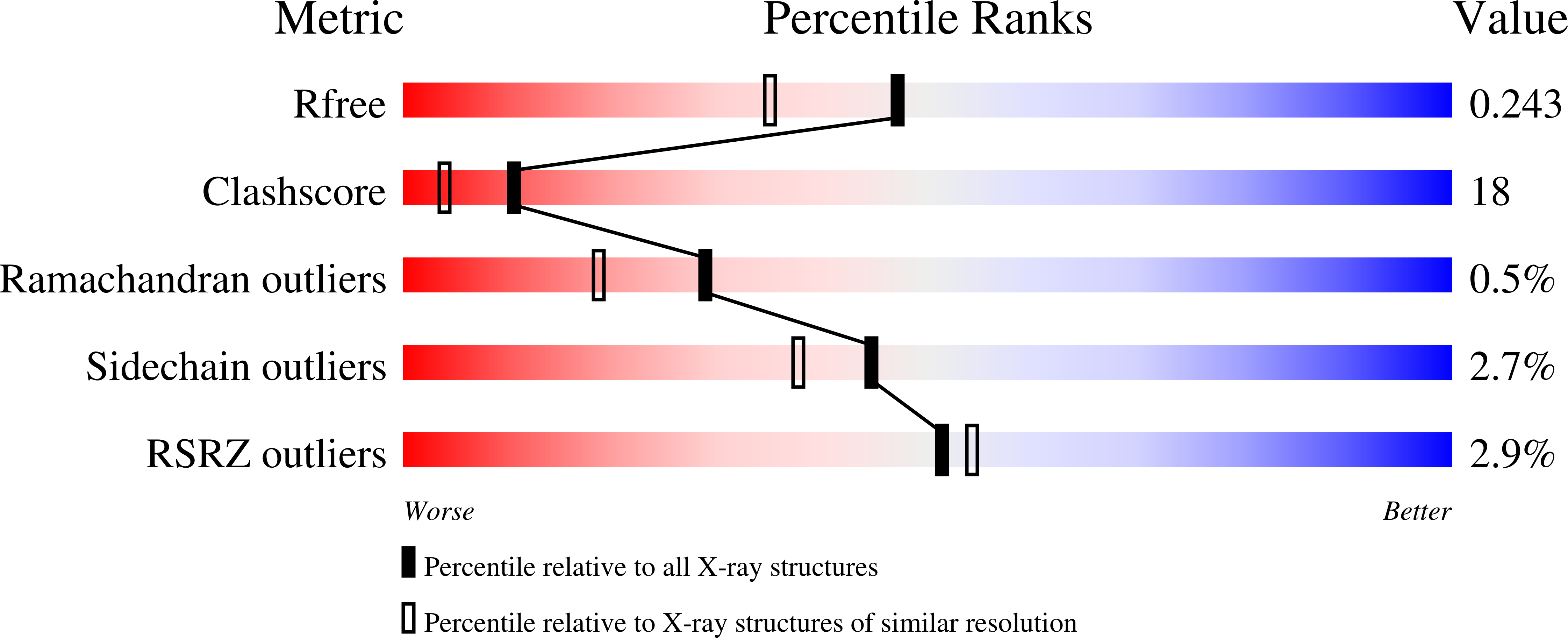
R-Value Free:
0.24
R-Value Work:
0.20
R-Value Observed:
0.22
Space Group:
P 6