3FH6 image
Deposition Date 2008-12-08
Release Date 2009-03-03
Last Version Date 2023-09-06
Entry Detail
PDB ID:
3FH6
Title:
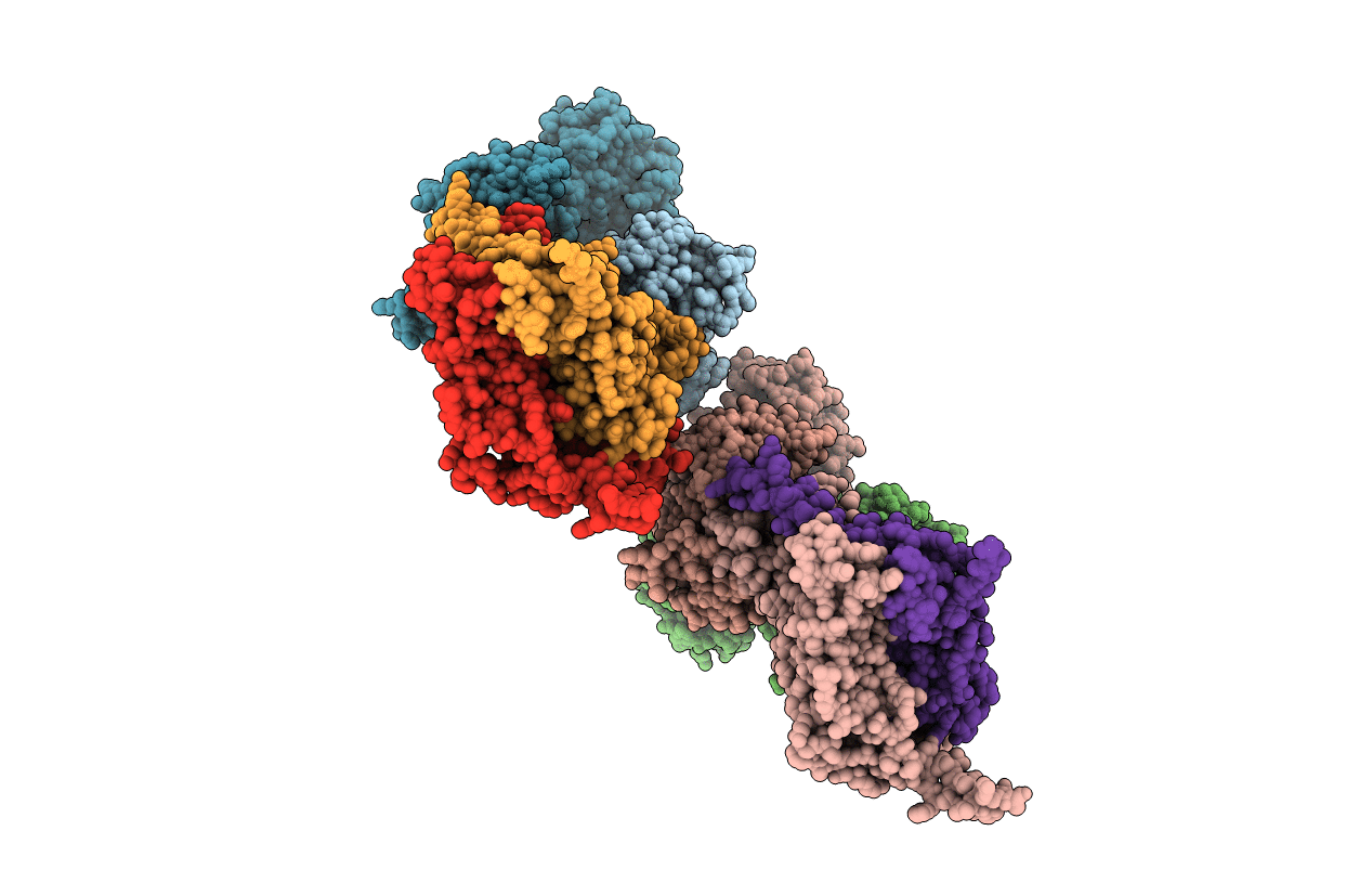
Crystal structure of the resting state maltose transporter from E. coli
Biological Source:
Source Organism:
Host Organism:
Method Details:
Experimental Method:
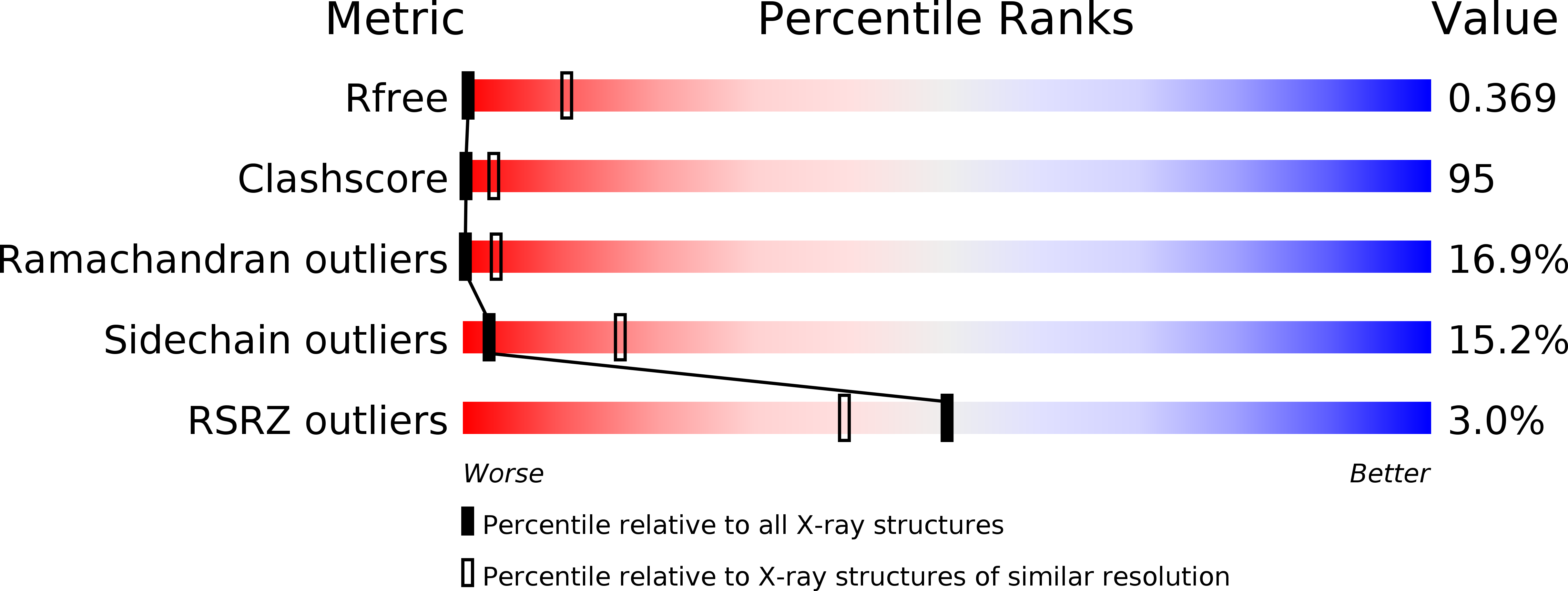
Resolution:
4.50 Å
R-Value Free:
0.36
R-Value Work:
0.34
Space Group:
I 2 2 2
Macromolecular Entities
Polymer Type:polypeptide(L)
Molecule:Maltose/maltodextrin import ATP-binding protein malK
Gene (Uniprot):malK
Chain IDs:C (auth: A), D (auth: B), G (auth: C), H (auth: D)
Chain Length:381
Number of Molecules:4
Biological Source:Escherichia coli
Polymer Type:polypeptide(L)
Molecule:Maltose transport system permease protein malF
Gene (Uniprot):malF
Chain IDs:A (auth: F), E (auth: H)
Chain Length:480
Number of Molecules:2
Biological Source:Escherichia coli
Polymer Type:polypeptide(L)
Molecule:Maltose transport system permease protein malG
Gene (Uniprot):malG
Chain IDs:B (auth: G), F (auth: I)
Chain Length:296
Number of Molecules:2
Biological Source:Escherichia coli
Ligand Molecules
Primary Citation
Alternating access in maltose transporter mediated by rigid-body rotations.
Mol.Cell 33 528 536 (2009)
PMID: 19250913 DOI: 10.1016/j.molcel.2009.01.035

Abstact

ATP-binding cassette transporters couple ATP hydrolysis to substrate translocation through an alternating access mechanism, but the nature of the conformational changes in a transport cycle remains elusive. Previously we reported the structure of the maltose transporter MalFGK(2) in an outward-facing conformation in which the transmembrane (TM) helices outline a substrate-binding pocket open toward the periplasmic surface and ATP is poised for hydrolysis along the closed nucleotide-binding dimer interface. Here we report the structure of the nucleotide-free maltose transporter in which the substrate binding pocket is only accessible from the cytoplasm and the nucleotide-binding interface is open. Comparison of the same transporter crystallized in two different conformations reveals that alternating access involves rigid-body rotations of the TM subdomains that are coupled to the closure and opening of the nucleotide-binding domain interface. The comparison also reveals that point mutations enabling binding protein-independent transport line dynamic interfaces in the TM region.

Legend

Protein

Chemical

Disease

Primary Citation of related structures