3FGS image
Deposition Date 2008-12-08
Release Date 2009-05-19
Last Version Date 2024-11-20
Entry Detail
PDB ID:
3FGS
Keywords:
Title:
Crystal structure of G65R/K206E double mutant of the N-lobe human transferrin
Biological Source:
Source Organism:
Homo sapiens (Taxon ID: 9606)
Host Organism:
Method Details:
Experimental Method:
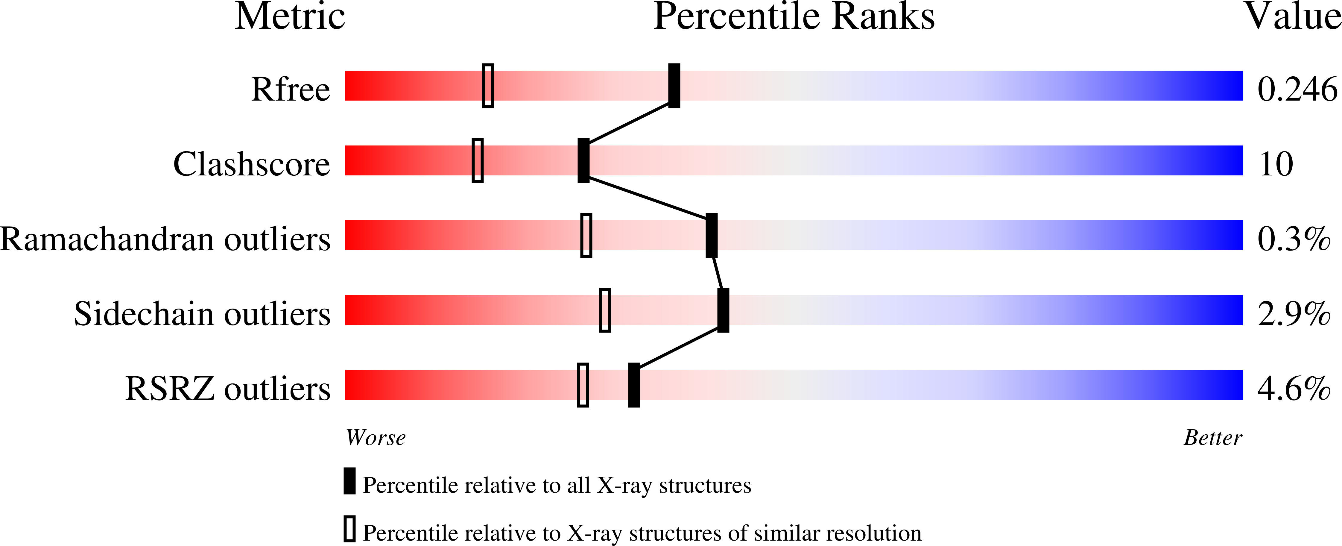
Resolution:
1.80 Å
R-Value Free:
0.24
R-Value Work:
0.21
Space Group:
P 21 21 21
Macromolecular Entities
Polymer Type:polypeptide(L)
Molecule:Serotransferrin
Gene (Uniprot):TF
Mutations:G65R, K206E
Chain IDs:A
Chain Length:337
Number of Molecules:1
Biological Source:Homo sapiens
Primary Citation
Structural and Functional Consequences of the Substitution of Glycine 65 with Arginine in the N-Lobe of Human Transferrin.
Biochemistry 48 1945 1953 (2009)
PMID: 19219998 DOI: 10.1021/bi802254x

Abstact

The G65R mutation in the N-lobe of human transferrin was created to mimic a naturally occurring variant (G394R) found in the homologous C-lobe. Because Gly65 is hydrogen-bonded to the iron-binding ligand Asp63, it comprises part of the second-shell hydrogen bond network surrounding the iron within the metal-binding cleft of the protein. Substitution with an arginine residue at this position disrupts the network, resulting in much more facile removal of iron from the G65R mutant. As shown by UV-vis and EPR spectroscopy, and by kinetic assays measuring the release of iron, the G65R mutant can exist in three forms. Two of the forms (yellow and pink in color) are interconvertible. The yellow form predominates in 1 M bicarbonate; the pink form is generated from the yellow form upon exchange into 1 M HEPES buffer (pH 7.4). The third form (also pink in color) is produced by the addition of Fe(3+)-(nitrilotriacetate)(2) to apo-G65R. Hydrogen-deuterium exchange experiments are consistent with all forms of the G65R mutant assuming a more open conformation. Additionally, mass spectrometric analysis reveals the presence of nitrilotriacetate in the third form. The inability to obtain crystals of the G65R mutant led to development of a novel crystallization strategy in which the G65R/K206E double mutation stabilizes a single closed pink conformer and captures Arg65 in a single position. Collectively, these studies highlight the importance of the hydrogen bond network in the cleft, as well as the inherent flexibility of the N-lobe which, although able to adapt to accommodate the large arginine substitution, exists in multiple conformations.

Legend

Protein

Chemical

Disease

Primary Citation of related structures