3FG2 image
Deposition Date 2008-12-04
Release Date 2009-09-01
Last Version Date 2023-11-01
Entry Detail
PDB ID:
3FG2
Keywords:
Title:
Crystal Structure of Ferredoxin Reductase for the CYP199A2 System from Rhodopseudomonas palustris
Biological Source:
Source Organism:
Host Organism:
Method Details:
Experimental Method:
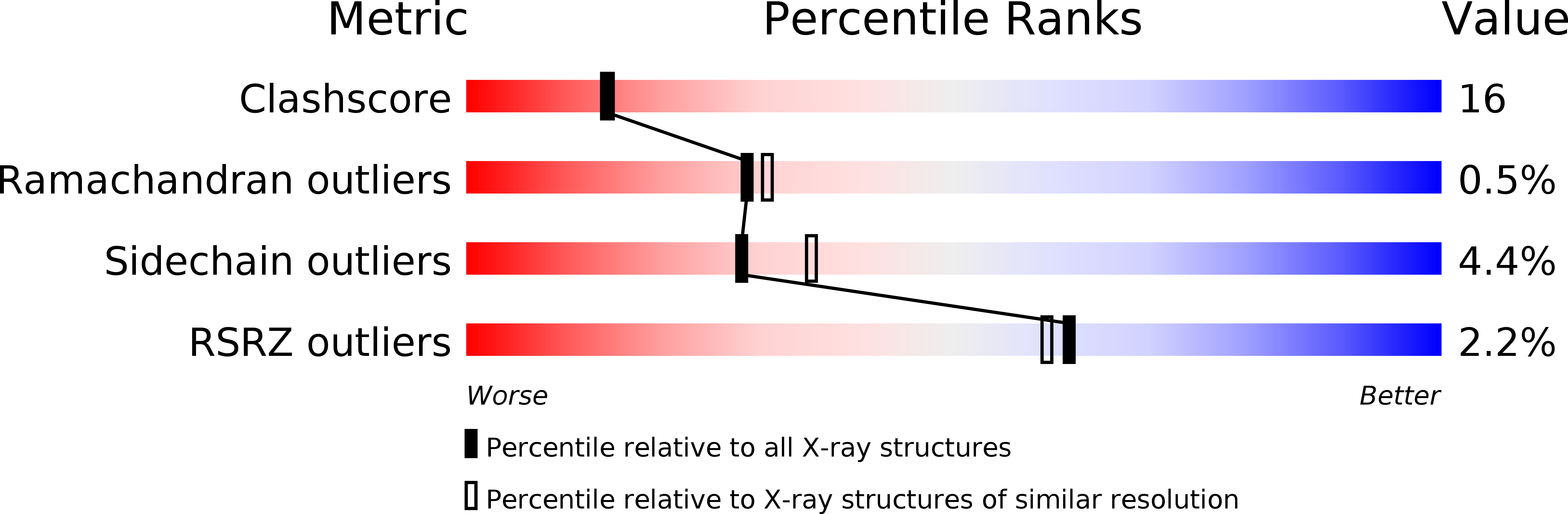
Resolution:
2.20 Å
R-Value Free:
0.25
R-Value Work:
0.21
Space Group:
P 32 2 1
Macromolecular Entities
Structures with similar UniProt ID
Protein Blast
Polymer Type:polypeptide(L)
Molecule:Putative rubredoxin reductase
Gene (Uniprot):TX73_019580
Chain IDs:A (auth: P)
Chain Length:404
Number of Molecules:1
Biological Source:Rhodopseudomonas palustris
Primary Citation
Crystal structure of a ferredoxin reductase for the CYP199A2 system from Rhodopseudomonas palustris
Proteins 77 867 880 (2009)
PMID: 19626710 DOI: 10.1002/prot.22510

Abstact

Cytochrome P450-199A2 from Rhodopseudomonas palustris oxidizes para-substituted benzoic acids and may play a role in lignin and aromatic acid degradation pathways in the bacterium. CYP199A2 has an associated [2Fe-2S] ferredoxin, palustrisredoxin (Pux) but not a ferredoxin reductase. A genome search identified the palustrisredoxin reductase (PuR) gene. PuR was produced in Escherichia coli and shown to be a flavin-dependent protein that supports efficient electron transfer from NADH to Pux, thus reconstituting CYP199A2 monooxygenase activity (k(cat) = 37.9 s(-1) with 4-methoxybenzoic acid). The reduction of Pux by PuR shows K(m) = 4.2 microM and k(cat) = 262 s(-1) in 50 mM Tris, pH 7.4. K(m) is increased to 154 microM in the presence of 200 mM KCl, indicating the importance of ionic interactions in PuR/Pux binding. The crystal structure of PuR has been determined at 2.2 A resolution and found to be closely related to that of other oxygenase-coupled NADH-dependent ferredoxin reductases. Residues on the surface that had been proposed to be involved in ferredoxin reductase-ferredoxin binding are conserved in PuR. However, Lys328 in PuR lies over the FAD isoalloxazine ring and, together with His11 and Gln41, render the electrostatic potential of the surface more positive and may account for the greater involvement of electrostatic interactions in ferredoxin binding by PuR. Consistent with these observations the K328G mutation weakened Pux binding and virtually eliminated the dependence of PuR/Pux binding on salt concentration, thus confirming that the FAD si side surface in the vicinity of Lys328 is the ferredoxin binding site.

Legend

Protein

Chemical

Disease

Primary Citation of related structures